Heart Development – Embryology Made Easy

Heart Tube

At the beginning of 4th week of development, heart is a continuous and valveless linear tube that resembles a chicken hung upside-down. It consists of 5 embryonic dilatation, that are destined to be the inflow and outflow tract and compartments of the hear without septum and valves.

heart tube mnemonic
Use the analogy of chicken hung upside-down:
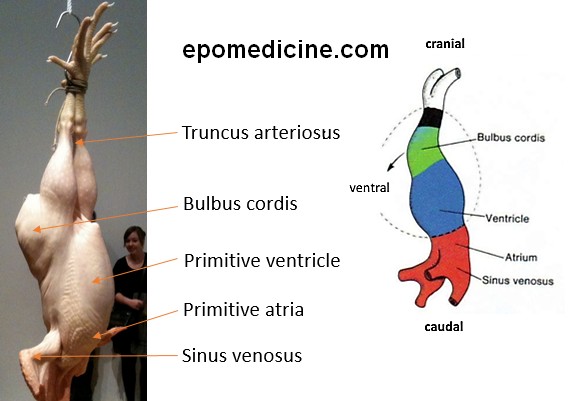
1. Legs: Outflow (truncus arteriosus); 2. Wings: Inflow (Sinus venosus); 3. Thigh: Bulbus cordis; 4. Breast: Primitive ventricle and primitive atria

From cranial to caudal, these dilations are:

  1. Truncus arteriosus (arteries)
    • Ascending aorta
    • Pulmonary trunk
    • Distal end dilates to form aortic sac which divides into right and left limbs. Each limb is connected to corresponding dorsal aorta through 6 aortic arches.
  2. Bulbus cordis
    • Smooth part of left (conus arterious) antd right ventricles (aortic vestibule)
  3. Primitive ventricle
    • Trabeculated part of left and right ventricles
  4. Primitive atrium
    • Trabeculated part of left and right atrium
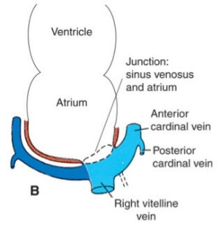
  5. Sinus venosus
    • Right – smooth part of right atrium (sinus venarum)
    • Left – coronary sinus
    • Oblique vein of left atrium

Smooth part of left atrium is formed by primitive pulmonary veins.

Crista terminalis: Junction of the trabeculated and smooth parts of the right atrium
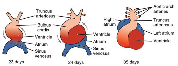
Dextral looping

  • The heart tube grows and starts to bend to the right at day 22 and is completed by the end of 4th week (day 28)
  • Retinoic acid is thought to act as a signal for antero-posterior values in heart development.
  • Looping depends on the laterality inducing genes.
  • Looping aligns the future atria, ventricles, inflow and outflow tract.
  • During this process, the proepicardial cells invest the outer layer of heart tube and eventually form epicardium  and coronary vasculature.
  • Here, we will use the analogy of inverted sit ups to imagine how the looping occurs and how it positions the embryonic dilations of heart tube.
heart looping mnemonic
Bulbus cordis is displaced inferiorly, ventrally and to the right.
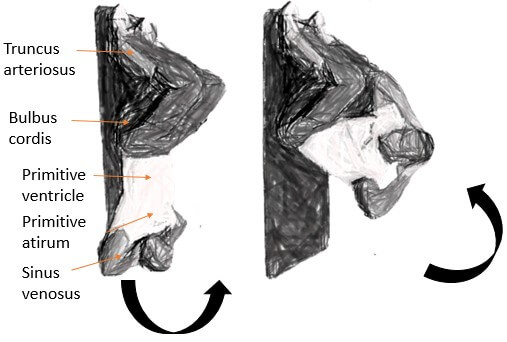
Ventricle goes to the left.
Atrium moves posteriorly and superiorly.
As the heart continues to grow and bend – bulbus and ventricles come to lie side by side and ventral to atrium.
heart looping
Actual heart looping
Septation of heart
Septation of heart

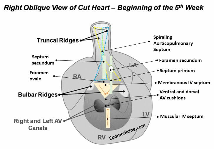
1. AP septum: Neural crest cells invade truncal and bulbar ridges leading to their growth in spiral fashion and formation of AP septum

2. Interatrial septum: Septum primum grows towards AV cushion and closes foramen primum but has foramen secundum in center. Septum secundum grows to the right and the gap between its upper and lower limb is foramen ovale.

3. AV septum: Ventral and dorsal AV cushion fuses to form right and left AV canal.

4. Interventricular spetum: Muscular IV septum grows from the floor of ventricle towards AV cushion and remaining gap is IV foramen. Membranous IV septum is formed by fusion of both bulbar ridges with dorsal AV cushion to close IV foramen.


sinus venosus development
Partial obliteration of left sinus venosus leading to left to right shunting and enlargement of right sinus venosus and shift of sinoatrial orifice to the right – sinus venosus opens only in right atrium.

Timeline of Other Important Events

Normal TimeDevelopmental EventsMalformations arising during period
Mid 3rd weekHorseshoe-shaped cardiac primordium appears.

  • Invaginating mesoderm cranial to prechordal plate
  • Wraps around the upper end of neural plate
  • Primary Heart Field (PHF) forms earlier and is lateral to Secondary Heart Field (SHF)

2 Endocardial tubes form on either side of neural plate.

  • Underlying foregut endoderm secretes VEGF leading to differentiation of PHF mesoderm into angioblastic cells.
Lethal mutations
20 daysBilateral Endocardial tubes fuse to form a single heart tube.

  • Fusion occurs cranio-caudally.
  • Due to lateral folding of the embryo.
  • Also due to lateral folding, heart tube is pinched off from primitive foregut and comes to lie ventral to it but remains connected to it by dorsal mesocardium
  • Coelomic cavity sourrounds the heart tube (future pericardial cavity)
  • NKX2.5 is the master gene for heart development.
Cardiac bifida (experimental)
Cardiac jelly appears.

  • Adjacent mesoderm called Mioepicardic plate gives rise to – developing myocardium and epicardium.
  • Between endocardium and mioepicardic plate, the plate secretes Extracellular Matrix – Cardiac Jelly
  • Cardiac jelly contains about 50 proteins including collagen, fibronectin, fibrillin, fibulin and growth factors.
Aortic arch is forming.

  • Connect heart tube to paired dorsal aorta.
22 daysHeart is looping into S shape.Dextrocardia

  • Heart lies on right side.
  • Dextrocardia can also be induced at an earlier time when laterality is established.
Heart begins to beat.
Dorsal mesocardium is breaking down.

  • Formation of transverse pericardial sinus.
Aortic arches I and II are forming.
24 daysAtria are beginning to bulge.
Right and left ventricles are likely to pump in series.
Outflow tract is distinguished from right ventricle.
Late week 4Sinus venosus is being incorporated into right atrium. Sinus venosus receives 3 sets of veins on either horns:

  1. From yolk sac: vitelline veins (future portal system)
  2. From chorionic villi: umbilical veins (placental)
  3. From cranial region and body of embryo: anterior and posterior cardinal veins via common cardinal vein (future caval system)
Venous inflow malformations
Endocardial cushions appear. Endocardial cushions are derived from:

  1. Endocardial epithelial cells that undergo Epithelial to Mesenchymal Transformation (EMT)
  2. Cardiac neural crest cells

Once, EMT has occured, both types of cells proliferate and invade the Cardiac Jelly, to form the endocardial cushions.

Sox9 plays an important role in endocardial cushion formation.

Persistent common atrioventricular canal


Septum primum appears from primitive atrial roof between right and left atria.Common atrium (Cor triloculare biventriculare):

  • Complete failure of septum primum and septum secondum to develop
Muscular interventricular septum is forming from the floor of primitive ventricle.

  • grows toward the fused AV cushions.
  • IV foramen is located between the free edge of the muscular IV septum and the fused AV cushions.
Common ventricle
Truncoconal ridges are forming.

  • 2 spiral (mesodermal) ridges grow from inner walls of truncus arteriosus on the opposite wall.
  • Similar ridges form in the bulbus cordis (conal or bulbar ridge) and become continuous with truncal ridges.
Persistent trucus arteriosus

  • Partial development of AP septum due to abnormal neural crest migration
  • One large vessel leaves the heart and receives blood from both the ventricles
  • Marked cyanosis due to R→L shunting
  • Often accompanied by membranous VSD
Aortic arch I is regressing.
Aortic arch III is forming.
Aortic arch IV is forming.
Early week 5Endocardial cushions are coming together, forming right and left atrioventricular canals.Persistent atrioventricular canal.
Sinoatrial orifice shifts to right:

  • Left horn of sinus venosus becomes partially obliterated (only Left umbilical vein is Left forming Ligamentum teres of Liver after obliteration of left common cardinal vein during 10th week)
  • Right horn of sinus enlarges due to Left to right shunting.
  • Right sinus horn becomes the inflow tract and opens only into the right atrim as the sinoatrial orifice shifts to right.
  • Right anterior cardinal vein becomes superior venacava.
  • Right vitelline vein becomes inferior venacava.
  • Right umbilical vein Regresses.
  • Remnant of left sinus venosus forms: oblique vein of left atrium and coronary sinus
Further growth of interatrial septum primum and muscular interventricular septum occurs.Muscular ventricular septal defects
Truncus arteriosus is dividing into aorta and pulmonary artery.

  • Neural crest cells and invade both the truncal ridges and the bulbar ridges.
  • The truncal and bulbar ridges grow and twist around each other in a spiral fashion and eventually fuse to form the AP septum.
  • The AP septum divides the truncus arteriosus and bulbus cordis into the aorta and pulmonary trunk.
Transposition of great vessels:

  • D-transposition (complete): nonspiral AP septum development due to abnormal neural crest migration → Aorta arises from right ventricle and pulmonary artery from left ventricle → Separate systemic and pulmonary circulation → Incompatible with life unless L to R shunt like VSD, ASD or Patent ductus arteriosus
  • L-transposition (corrected): D-transposition with inversion of the ventricles such that anatomical left ventricle lies on right and vice versa.

Tetralogy of Fallot:

  • Skewed development of AP septum due to abnormal neural crest migration leading to overriding of aorta and pulmonary stenosis (with VSD and RVH) leading to R→L shunting and cyanosis.
Atrioventricular bundle is forming; there is possible neurogenic control of heartbeat.
Pulmonary veins are being incorporated into the left atrium.

  • Trabeculated tissue is displaced anteriorly and laterally which then form the left atrial appendage.
Aberrant pulmonary drainage
Aortic arches I and II have regressed.

  • Aortic arch I remnant forms maxillary artery.
  • Aortic arch II remnant forms stapedial and hyoidal arteries.
Aortic arches III and IV have formed.

  • Aortic arch III – proximal part forms common carotid arteries; distal part forms internal carotid arteries
  • Aortic arch IV – left forms mid portion of arch of aorta (proximal by aortic sac and distal by dorsal aorta); right forms proximal part of right subclavian artery (distal by right dorsal artery)
 Aortic arch VI is forming.

  • Proximal part – proximal part of right and left pulmonary artery
  • Distal part – right disappears; left forms ductus arteriosus (patency maintained by placenta derive PGs)
  • Distal part of pulmonary arteries develop from the buds of aortic arch VI, which grow towards developing lungs.
Late week 5 to Early week 6Endocardial cushions fuse.

  • The atrio-ventricular canal is divided by the fusion of the dorsal and ventral AV cushions.
  • The formation and subsequent remodeling of the AV cushions is retinoic acid-dependent, so disruption of retinoid signaling often produces AV canal defects
 Persistent common AV canal:

  • Has common AV valve
  • L→R shunting of atrial blood leads to enlarged right atrium and ventricle.
  • Mitral valve regurgitation leads to enlarged left atrium and ventricle.
Interatrial foramen secundum is forming in septum primum.
Interatrial septum primum is almost contacting endocardial cushions (for closure of foramen primum).Low atrial septal defects
Membranous part of interventricular septum starts to form.

  • forms from fusion of: right bulbar ridge, left bulbar ridge and AV cushion
Membranous ventricular septal defect

  • L→R shunting leading to Pulmonary hypertension and later revershal of shunt (Eisenmenger’s syndrome)
Semilunar valves begin to form.

  • formed via cavitation of truncoconal ridge tissue to form three triangular valve leaflets in each of the outflow vessel in a highly stereotypical pattern:
  • pulmonary semilunar valve develops three cusps: left, right, and anterior
  • aortic semilunar valve also develops three cusps: left, right and posterior
Aortic and pulmonary vascular stenosis
Late week 6Interatrial foramen secundum is large.High atrial septal defects
Interatrial septum secundum starts to form.

  • Locate to the right of septum primum
  • Between the upper and lower limb of septum secundum is foramen ovale.
Foramen secundum defect: Excessive resorption of septum primum or secundum or both. It may be tolerated for a long time.
Atrioventricular valves and papillary muscles are forming.Ebstein’s anomaly:

  • Low insertion of tricuspid valve into the right ventricle
  • Tricuspid regurgitation
  • Atrialization of upper large portion of ventricle
  • Only small lower portion of right ventricle is functional

Tricuspid atresia (hypoplastic right heart):

  • Insufficient AV cushion to form tricuspid valve
  • No communication between right atrium and ventricle
  • Marked cyanosis, Patent foramen ovale, IV septum defect, underdeveloped right ventricle
Interventricular septum is almost complete.Membranous ventricular septal defects
Coronary circulation is being established.
8-9 weeksMembranous part of interventricular septum is complete.

  • Closure of IV foramen
Membranous ventricular septal defects


Write your Viewpoint đź’¬

Your email address will not be published. Required fields are marked *

This site uses Akismet to reduce spam. Learn how your comment data is processed.