Ectrodactyly is an autosomal dominant ectodermal dysplasia presenting as bilateral congenital malformed hands and feet [1]. It affects about 1 in 90,000 births with males and females equally as likely to be affected.
It is characterized by transverse terminal aphalangia or partial to total absence of the distal segments of fingers. It may involve one or more digits or the full hand and even part of the upper arm. More severe manifestations are hemimelia or amelia. All these abnormalities are considered to represent various degrees of severity of the same anomaly and may be due to an intrauterine vascular occlusion or insufficiency [2]. These different forms are connected with a different genetic mutation. Type I, the most frequent form has been found to be a mutation on chromosome 7 in a region that contains two homeobox genes, DLX5 and DLX6.
Usually this is characterized as the split hand/foot deformity due to the absence of the third digit, with clefting into the proximal portion of the hand or foot and syndactyly of remaining digits on each side of the cleft. The hand resembles a lobster claw [3]. The association of ectrodactyly with cleft lip and palate was originally described by Cockayne [4]. It was known as Ectrodactyly-Ectodermal Dysplasia-Cleft lip/palate syndrome (EEC syndrome) [5].
The case:
A female baby was born in Hospital. The primigravidae mother had no significant medical history. There was no history of consanguinity or any other relevant family history. She had uneventful antenatal period and had received all the supplements. There was no gestational diabetes mellitus, pregnancy induced hypertension. Antenatal ultrasound at local health center showed no congenital abnormalities. After a term pregnancy of 38 weeks, caesarian section was performed for oligohydramnios with amniotic fluid index of 2. Birth weight was 2.4 kilograms. No resuscitation was required.
Physical examination revealed the following:
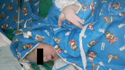
Both her hands were showing lengthening and broadening of the digits. There was a medial cleft in the metacarpals, dividing the hand into two portions. Syndactyly of the remaining fingers was seen. The growth of the digits was more as compared to other body parts. The nails of the affected fingers were maldeveloped . Her legs were normal. Systemic examination of the patient did not reveal any other anomaly. Abdominal ultrasonography did not show any abnormality. Echocardiogram revealed double outlet right ventricle. Hair and teeth were normal and there were no other congenital malformations.
Discussion:
Ectrodactyly is a rare autosomal dominant ectodermal dysplasia. It sometimes may be associated with other ectodermal defects. The most common clinical manifestations of EEC syndrome are ectodermal dysplasia, ectrodactyly, cleft lip/palate and tear duct anomalies. The expression of this may be quite variable with reduced penetrance also. In a review of 230 published cases by Roelfsema et al. [6], ectrodactyly was found in 84%, ectodermal dysplasia in 77%, clefting in 68% and anomalies of lacrimal ducts in 59%. Urogenital defects were reported in 52%. Isolated cases were more severely affected than familial cases [6]. In the present case she only showed lobster claw with double outlet right ventricle. The parents were offered genetic counseling and the mode of inheritance explained. Since ectrodactyly is an autosomal dominant disorder, there are 50% chances of recurrence for the future pregnancies. Genetic studies using mutation analysis was explained to the patient but the patient opted out, as it was very expensive and not available in Nepal. In the present case, the parents left against medical advice.
References
1. Kalla G, Garg A. Ectrodactyly. Indian J Dermatol Venereol Leprol. 2002;68:152–153.
2. Deborah K. The dysostoses. In: Rimoin DL, Connor JM, Pyeritz RE and Korf BR, editors. Principles and practice of medical genetics. 4th ed. London: Churchill Livingstone. 2002; pp. 4170–4171.
3. Jung KR, Jeong C, Jong SC. Ectrodactyly. Korean J Radiol. 2003;4(4):243–251. doi: 10.3348/kjr.2003.4.4.243.
4. Cockayne EA. Cleft palate-lip, hair lip, docrocystitis and cleft hand and foot. Biometrika. 1936;26:60–63.
5. Pries C, Mittleman D, Miller M, et al. The EEC syndrome. Am J Dis Child. 1974;127:840–844.
6. Roelfsema NM, Cobben JM. The EEC syndrome: a literature study. Clin Dysmorphol. 1996;2:115–127.
Author:

Dr. Nischal Maskey
MD Pediatrics (Institute of Medicine)
Thank you for Very nice information, is there possibility of surgical correction of such type of anomaly
If indicated, the operative options are thumb, thumb web space, and central cleft reconstruction. If the cleft hand doesn’t have progressive deformities, surgery can take place when the child is 1 or 2 years old.