Skip to content
Epomedicine

Mnemonics, Simplified Concepts & Thoughts

Epomedicine

Mnemonics, Simplified Concepts & Thoughts

renal angle tenderness

Costovertebral or Renal angle tenderness

Epomedicine, Jul 26, 2019Jul 26, 2019

Synonyms: Murphy’s kidney punch, CVA tenderness (CVAT)

Anatomy of Costovertebral angle or Renal angle:

Costovertebral angle is formed by the junction of the 12th, or lowermost, rib with the paravertebral muscles, which run parallel to and on both sides of the vertebral column.

Eliciting costovertebral angle tenderness:

costovertebral angle tenderness

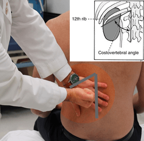
Position of patient: Sitting or Prone

It is performed by striking the fist of one hand against the dorsal surface of the other hand, which is placed flat along the posterior CVA margin. Normally, percussion in CVA should not elicit tenderness.

Causes of costovertebral angle tenderness:

  1. Acute pyelonephritis
  2. Calculi
  3. Renal artery occlusion
  4. Perinephric abscess

References:

  1. Nursing Know-how: Evaluating signs & symptoms – Lippincott Williams & Wilkins
  2. Varney’s Midwifery – By Helen Varney, Helen Varney Burst, Jan M. Kriebs, Carolyn L. Gegor
  3. Emergency and Trauma Care for Nurses and Paramedics – By Kate Curtis, Clair Ramsden
  • Facebook
  • Twitter
Clinical Skills and Approaches Clinical examinationGeneral SurgeryInternal medicinePediatricsRenal and Electroloyte

Post navigation

Previous post
Next post

Related Posts

Clinical Skills and Approaches

FARES method of Shoulder Reduction

Nov 30, 2019Nov 30, 2019

Indication: Anterior shoulder dislocation Position of patient: Supine Position of physician: Standing on the side of the dislocated shoulder Steps: Physician holds the wrist of the patient with both the arms keeping elbow of the patient extended and forearm in neutral position Arm is slowly abducted in brief oscillating movement…

Read More
Clinical Skills and Approaches

Common mistakes in Per Abdominal examination

Jul 31, 2020Jul 31, 2020

1. Forgetting to Expose abdomen adequately: Before examination, patient should ideally be exposed from the nipples to mid thigh. Failure to do so may lead to missed findings during examination e.g. Hernia 2. Abdominal symmetry and movement: Abdominal symmetry and movement should be examined tangentially and from leg end. Comment…

Read More
Clinical Skills and Approaches hand examination

Examination of Hands

Mar 18, 2015

History: Hand dominance and occupation? Injury? Pain? Paresthesia? Impaired function? Swelling? Position: Place the patient’s hands on pillow Look: SEATS a. Shape or Deformity: – Wrist: Radial deviation: RA Ulnar deviation and flexion deformity: Spastic hemiplegia (CP) Wrist drop (also finger drop): Radial nerve injury Prominent dorsal ulnar and radial…

Read More

Leave a Reply Cancel reply

Your email address will not be published. Required fields are marked *

This site uses Akismet to reduce spam. Learn how your comment data is processed.

Pre-clinical (Basic Sciences)

Anatomy

Biochemistry

Community medicine (PSM)

Embryology

Microbiology

Pathology

Pharmacology

Physiology

Clinical Sciences

Anesthesia

Dermatology

Emergency medicine

Forensic

Internal medicine

Gynecology & Obstetrics

Oncology

Ophthalmology

Orthopedics

Otorhinolaryngology (ENT)

Pediatrics

Psychiatry

Radiology

Surgery

RSS Ask Epomedicine

  • What to study for Clinical examination in Orthopedics?
  • What is the mechanism of AVNRT?

Epomedicine weekly

  • About Epomedicine
  • Contact Us
  • Author Guidelines
  • Submit Article
  • Editorial Board
  • USMLE
  • MRCS
  • Thesis
©2026 Epomedicine | WordPress Theme by SuperbThemes