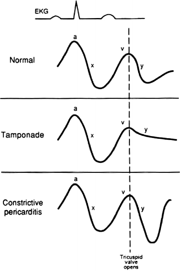
Jugular venous pulse (JVP) waveforms
- a – Atrial contraction (RA)
- c – Closure and Curving of tricuspid valve into RA
- x – atrial relaXation
- v – Venous filling of right atrium (RA)
- y – atrial emptYing
Distinguishing JVP from Internal carotid pulsation
Mnemonic: POLICE
- Palpation: Non-palpable
- Occlussion: Readily occludable
- Location: Between 2 heads of sternocleidomastoid, lateral to carotid
- Inspiration: Drops with inspiration
- Contour: Biphasic waveform
- Erect position: Drops when sitting erect
Abnormal JVP waveforms
Prominent “a” wave: Pressure of atria peaked
- Tricuspid stenosis
- Pulmonary hypertension
Cannon “a” wave: Contraction of atria against closed tricuspid valve
- Complete heart block
- Junctional rhythm
“a” wave fallen (absent): Atrial fibrillation
Large “v” wave: Vomit (backflow) of blood in right atrium
- Tricuspid regurgitation
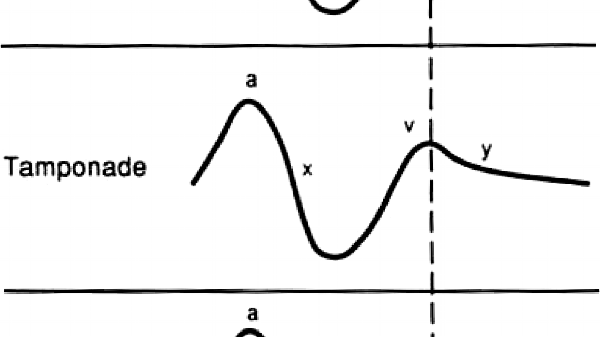
PaY TaX:
- Sharp “y” descent: Constrictive pericarditis (Frierdrich’s sign)
- Sharp “x” descent: Tamponade and Constrictive pericarditis
Slow “y” descent”: Slow emptYing of RA
- Tricuspid stenosis

He is the section editor of Orthopedics in Epomedicine. He searches for and share simpler ways to make complicated medical topics simple. He also loves writing poetry, listening and playing music. He is currently pursuing Fellowship in Hip, Pelvi-acetabulum and Arthroplasty at B&B Hospital.