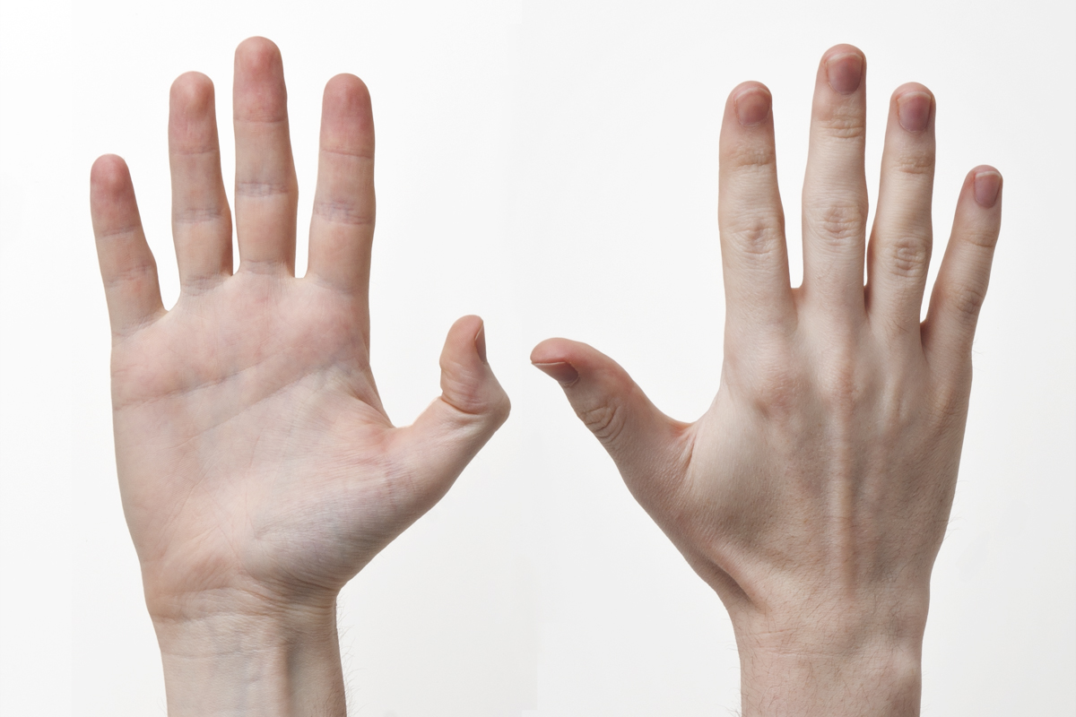
Look
Examine SEADS with palms facing upwards and downwards:
- Nails:
- Subungual exostosis
- Glomus tumor (bluish tinge)
- Ganglion cyst at nail bed (grooved nail)
- Psoriatic arthropathy (pitting and onycholysis)
- Swellings:
- Volar:
- Inclusion dermoid cysts
- Ganglion of A1 pulley (Pearl ganglion)
- GCT of flexor sheath (usually in relation to proximal phalanx)
- Dorsal:
- DIP: Heberden’s node, Mucous cyst
- PIP: Bouchard’s node, Garrod’s pad
- MCP: Rheumatoid nodule
- CMC (index and middle finger): Carpal boss
- Volar:
- Attitude:
- Palm upwards: Gradual cascade of fingers (increasing flexion in joints from index to little finger)
- Palm downwards: Gradual cascade of fingers (increasing extension in joints from little to index finger)
- Tenodesis effect of wrist movement
- Puckering and ridging/pitting and nodules of skin in the palm sometimes extending into finger: Dupuytren’s contracture
- Deformity: Look for rheumatoid hand deformities, clawing, angular and rotational deformities
Interrupted finger cascade = Tendon divided or stuck
Normal finger cascade but absent active motion = Nerve injury
Feel
Examine TEST CA
- Anhidrosis: Nerve damage
- Lump: Full examination of lump
- Nodule: Look for “triggering effect”
- Cords: Passively extend fingers and palpate for possible cords in palm (Dupuytren’s)
- Autonomous sensory areas:
- Tip of index finger: Median nerve
- Base of thenar area: Palmar cutaneous branch of median nerve
- Tip of little finger: Ulnar nerve
- Dorsum of 5th metacarpal: Dorsal cutaneous branch of ulnar nerve
- Dorsal 1st webspace: Radial nerve
Move
Assess active and then passive ROM.
- Composite movement: Look for extension and flexion of fingers (fingertip to palm distance can be used as a quick measure of movement loss)
- Finger joints:
- DIP: 0-80 degrees
- PIP: 0-100 degrees
- MCP: 0-40 degrees hyperextension to 90 degrees flexion
- Thumb:
- Extension (sideways movement in the plane of the palm)
- Abduction (upward movement at right angle to the palm)
- Adduction (pressing against the palm)
- Flexion (sideways movement across the palm)
- Opposition (touching the tips of the fingers)
- Retropulsion (lifting the thumb backwards behind the plane of the hand)
Muscles and Tendons
- FDP: Immobilize PIP in extension and ask to flex DIP joint
- FDS of non-index fingers: Grasp all fingers in extension (FDP with common belly is restricted), except the one being tested and ask to flex the isolated finger
- FDS of index finger: Ask to pinch hard with DIP in extension and PIP in full flexion (FDP of index finger has a separate belly)
- FPL: Immobilize thumb MCP in extension and ask to flex IP joint
- EPL: Check retropulsion of thumb
- Long extensors: Ask to extend MCP joint
- EIP (found on ulnar side of EDC): Ask to flex MCP joints of all other fingers (defunction EDC), and extend index finger actively; can be used for tendon transfer
- Intrinsic mucles: Ask to make ‘duckbill’ position, i.e. Flex MCP joints and extend IP joints simultaneously
- PL (Schaeffer’s test): Ask to oppose thumb to little finger and flex the wrist (useful for tendon transfers); absent in 15%
Grip
- Pick a pin: Precision grip
- Hold a sheet of paper: Pinch grip
- Hold a pen: Tripod/chuck grip
- Hold a key (side to end): Key grip
- Hold my curled fingers: Hook group
- Grip my forearm: Grasp/span
- Handshake: Power grip
- Grip strength: Ask patient to squeeze partially inflated sphygmomanometer cuff (20 mmHg) – normally a pressure of 150 mmHg can be achieved easily
Special tests
1. Allen’s test: Patient clenches fingers several times before making a fist (exsanguination), then examiner occludes radial and ulnar artery simultaneously with fingers. Ask patient to release fist (must stay blanched, else the occlusion pressure is not adequate). Release compressed artery one at a time to test that artery. Normal color must return within 5-15 seconds.
- Digital Allen’s test: Similar to Allen’s test but digital arteries are occluded.
2. Bunnel’s test: Assess passive PIP joint flexion in 2 positions:
- In MCP extension (intrinsics in tension) < In MCP flexion (instrinsics relaxed): Intrinsic tightness
- In MCP extension (extrinsics relaxed) > In MCP flexion (extrinsics tightened): Extrinsic tightness
- Limited in both MCP extension and flexion: Capsular tightness
3. Bouvier’s test: In a claw hand, correct MCP hyperextension – if patient is able to extend IP joints (tenodesis working), then the clawing is flexible.
4. Elson test: Ask to place hand flat on table with PIP joint flexed over the edge of table. Ask to actively extend finger against resistance:
- Intact central slip: Strong PIP extension (central slip works) and Relaxed DIP (lateral bands will remain slack due to tethering of extensor mechanism by central slip)
- Ruptured central slip: Weak PIP extension (central slip defunctions) and Hyperextension and stiffness of DIP (lateral band tensioning)
5. Test for ORL tightness: Assess passively flexion of DIP in 2 positions:
- In PIP flexion (relaxes ORL) > In PIP extension: ORL tightness
- In PIP flexion = In PIP extension: DIP joint contracture
6. 1st MCP valgus stress test:
- Instability in neutral (0 degrees): Injury to proper and accessory UCL and/or volar plate
- Instability in 30 degrees flexion: Injury to proper UCL
Examination of Peripheral Nerves will be discussed separately.