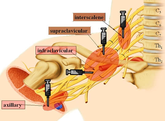
Brachial plexus is sub-divided from proximal to distal into: Roots, Trunks, Divisions, Cords, Branches. This can be easily remembered with a mnemonic: Randy Travis Drinks Cold Beer. Approaches for Brachial Plexus Block Basically, there are 4 approaches to the brachial plexus block at different levels as described in the mnemonic…
Category: PGMEE, MRCS, USMLE, MBBS, MD/MS
Medical knowledge in bullet points with understandable language, simplified images and graspable mnemonics.
PGMEE, MRCS, USMLE, MBBS, MD/MS
Local Anesthetics Mnemonic
All Local anesthetics contain suffix “-caine”. Local Anesthetics (LA) can be classified as: Esters and Amides. Esters vs Amides A mnemonic device is that the names of amides contain 2 “i”s compared with only 1 “i” seen in esters. Remember: One-eyed ester or Amide word has an “i” in it…
PGMEE, MRCS, USMLE, MBBS, MD/MS
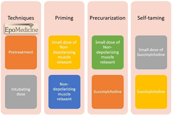
Priming, Precurarization and Self taming
Priming Administration of a small sub-paralyzing dose of non-depolarizing muscular blocking agent (usually 10% of the intubating dose) is given 2-4 minutes before administering a 2nd large dose for tracheal intubation to accelerate the onset of non-depolarizing NM blockade by 30-60 seconds. Mechanism and Concept of Priming 2 theories have…
PGMEE, MRCS, USMLE, MBBS, MD/MS
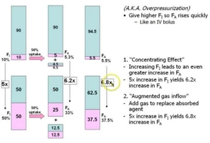
Concentration effect, Second gas effect and Diffusion hypoxia
Concentration effect The higher the concentration of an inhaled anesthetic, the faster the alveolar concentration approaches the inhaled concentration. This is referred to as concentration effect and is clinically significant only in cases where gases are administered in high concentration: Nitrous oxide Xenon Ostwald’s blood gas solubility coefficient: ratio of…
PGMEE, MRCS, USMLE, MBBS, MD/MS
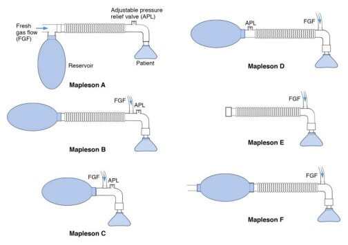
Mapleson Breathing Circuit Made Easy
Anatomy of Mapleson Breathing Circuit Basically, a mapleson breathing circuit consists of following parts: 1. Face mask (towards patient end) 2. Reservoir bag (towards operator end) Accommodates fresh gas flow during expiration acting as a reservoir available for the following inspiration. Acts as a monitor of patient’s ventilatory pattern during…
PGMEE, MRCS, USMLE, MBBS, MD/MS
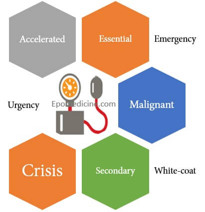
Terminologies of Hypertension
There are various terminologies used to describe hypertension which may overlap and are a source of confusion to the medical students and health professionals. Essential or Primary or Idiopathic hypertension Hypertension in which secondary causes have been excluded. Identifiable etiologic factors of essential hypertension: Obesity Insulin resistance High alcohol intake…
PGMEE, MRCS, USMLE, MBBS, MD/MS
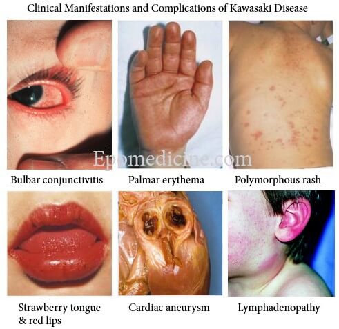
Kawasaki Disease – Diagnostic Criteria Mnemonic
The diagnostic criteria of Kawasaki Disease can be remembered using a mnemonic – “FEBRILE“. Fever: >5 days plus ≥4 of the following Enathem: Lips: Erythema, fissuring or crusting Oropharynx: Diffuse injection Tongue: Strawberry tongue Bulbar conjunctivitis: Bilateral, painless and non-exudative Rash: Polymorphous rash Internal organ involvement (not the part of criteria)…
PGMEE, MRCS, USMLE, MBBS, MD/MS
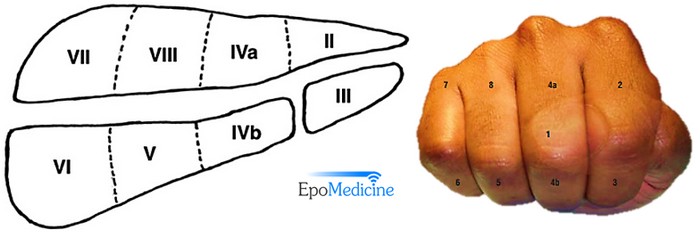
Liver Segments Explained with Mnemonic
Couniaud divided liver into 8 functional segments, each of which is supplied by it’s own portal triad (composed of a portal vein, hepatic artery and a bile duct). Hepatic veins divide the liver in saggital plane: 1. Middle hepatic vein: Divides the liver into right and left functional lobe. 2….