Couniaud divided liver into 8 functional segments, each of which is supplied by it’s own portal triad (composed of a portal vein, hepatic artery and a bile duct).
Hepatic veins divide the liver in saggital plane:
1. Middle hepatic vein: Divides the liver into right and left functional lobe.
- Cantle’s line: run from middle of gall bladder fossa anterior to Inferior venacava posteriorly
- Left lobe = Segment I-IV (Segment I is separately the caudate lobe)
- Right lobe = Segment V-VIII
2. Left hepatic vein: Divides left lobe into lateral and medial segments.
- Lateral segment = Segment II and III
- Medial segment = Segment IV
Falciform ligament: left hepatic vein is located slightly left to the left hepatic vein; hence, falciform ligament roughly divides liver into right and left lobe.
3. Right hepatic vein: Divides right lobe into anterior and posterior segments.
- Anterior segment = Segment V and VIII
- Posterior segment = Segment VI and VII
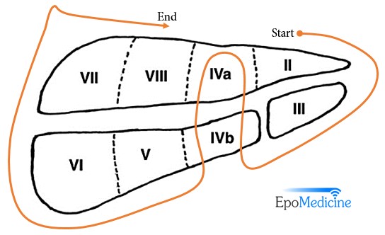
Portal vein divides the liver in transverse plane:
- Upper segment = Segment II, IVa, VIII, VII
- Lower segment = Segment III, IVb, V, VI
How to remember the orientation of Couniaud Liver Segments?
We have a handy hand mnemonic for this purpose.1
Also, remember that the segments are numbered in a clock-wise fashion.
With the right hand, make a fist while tucking the thumb behind the remainder of the fingers.
- Tucked in thumb = Segment I (Caudate Lobe)
- Line of Proximal Interphalangeal (PIP) joints = Plane of portal vein, separating liver into upper and lower segments.
- Interdigitary spaces = Intersegmental planes (hepatic fissures – left, middle and right) in which corresponding hepatic veins run and divide liver sagitally
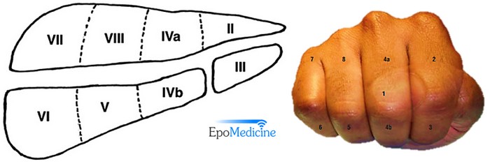
For the remaining 7 segmets, go in a clockwise direction starting from the lateralmost upper left part, i.e. Proximal phalax of 1st finger.
- Proximal phalanx of 1st finger = Left superior lateral segment = Segment II
- Distal phalanx of 1st finger = Left inferior lateral segment = Segment III
- 2nd finger = Left medial segment = Segment IV (Quadrate lobe)
- Proximal phalanx = Segment IVa
- Distal phalanx = Segment IVb
- Distal phalanx of 3rd finger = Right inferior anterior segment = Segment V
- Distal phalanx of 4th finger = Right inferior posterior segment = Segment VI
- Proximal phalanx of 4th finger = Right superior posterior segment = Segment VII
- Proximal phalanx of 3rd finger = Right superior anterior segment = Segment VIII
Segmental Anatomy of Liver
Portal vein:
The left portal vein supplies Couinaud segments I, II, III, and IV.
The right portal vein subdivides into anterior and posterior branches.
- Anterior branch: supplies segments V and VIII
- Posterior branch: supplies segments VI and VII.
Hepatic vein:
Right hepatic vein: drains all of segment VI and VII and some of segments V and VIII.
Left hepatic vein: drains segments II and III and IV.
Middle hepatic vein: drains segment IV, V and VIII.
Intrahepatic bile ducts:
Segment II-IV: Left hepatic duct
Segment V and VIII: Right anterior hepatic duct
Segment VI and VII: Right posterior hepatic duct
Caudate lobe Segment I):
The left lobe is supplied or drained by left branches from porta hepatis components and right from right. The caudate lobe is anatomically different from other lobes:
- It hase direct connections to the IVC through hepatic veins
- It may be supplied by both right and left branches of the portal vein.
- It is drained drained by both right and left hepatic ducts.
Caudate lobe is the lobe that is not affected in Budd-Chiari syndrome and cirrhosis of liver because of it’s direct drainage to IVC. For further information about liver diseases and types of diagnostic tests for liver conditions you can visit fibronostics.com.
Hepatectomy and Sectionectomy
Right hepatectomy = Segment V-VIII (± segment I)
Extended right hepatectomy or Right trisectionectomy = Right hepatectomy + Segment IV
Left hepatectomy = Segment II-IV (± segment I)
Extended left hepatectomy or Left trisectionectomy = Left hepatectomy + Segment V and VIII
Right posterior sectionectomy = segment VI and VII
Right anterior sectionectomy = segment V and VIII
Left medial sectionectomy = segment IV
Left lateral sectionectomy = segment II and III

He is the section editor of Orthopedics in Epomedicine. He searches for and share simpler ways to make complicated medical topics simple. He also loves writing poetry, listening and playing music. He is currently pursuing Fellowship in Hip, Pelvi-acetabulum and Arthroplasty at B&B Hospital.
Thank you very much! I appreciate your efforts! Blessings.