Skip to content
Epomedicine

Mnemonics, Simplified Concepts & Thoughts

Epomedicine

Mnemonics, Simplified Concepts & Thoughts

maxillary artery

Maxillary Artery : Mnemonic

Epomedicine, Oct 16, 2017Aug 23, 2023

Origin of Maxillary artery: Terminal branch of External Carotid Artery (ECA)

Derived from: 1st Arch

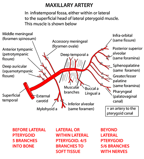
Divisions of Maxillary artery: 3 parts by lateral pterygoid

  1. 1st part (Mandibular part): winds around deep to neck of mandible
  2. 2nd part (Pterygoid part): travels between 2 heads of lateral pterygoid
  3. 3rd part (Pterygopalatine part): enters pterygopalatine fossa containing pterygopalatine galnglion
maxillary artery branches
Henry Vandyke Carter, Public domain, via Wikimedia Commons

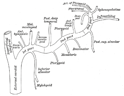
Branches of Maxillary artery

Remember:

  1. Each of the 3 divisions gives off 5 branches.
  2. Mandibular artery, i.e. Inferior alveolar artery is a branch of maxillary artery
  3. 2nd part: Branches supply muscles of mastication and do not cross through foramina in bones (all branches from 1st and 3rd part do cross)

Branches from 1st part

Mnemonic: Mandibular part gives off branches to 5 “M“s and the branches can be remembered as “DAMIA“

  1. Meatus (External auditory meatus) – Deep auricular
  2. Membrane (tympanic membrane) – Anterior tympanic
  3. Meninges and mater (duramater) – Medial meningeal
  4. Mandible – Inferior alveolar
  5. Meckel’s cave – Accessory meningeal

Clinical significance: Middle meningeal artery (MMA) injury results in Extradural hematoma (EDH). Auriculotemporal nerve wraps around it.

Branches from the 2nd part

Mnemonic: They supply muscles of mastication which are also derivatives of the 1st arch.

  1. Posterior deep temporal artery: Temporalis
  2. Pterygoid artery: Lateral and medial pterygoids
  3. Masseteric artery: Masseter
  4. Buccinator artery: Buccinator
  5. Anterior deep temporal artery: Temporalis

Branches from the 3rd part

Mnemonic: Remember the 5 “P“s

  1. Posterior superior alveolar artery: Maxilla
  2. Pterygoid canal artery: Upper part of pharynx and tympanic cavity
  3. Palatine – Descending palatine artery: Gives off greater and lesser palatine artery and supplies hard and soft palate respectively
  4. Palatine – Sphenopalatine or Nasopalatine artery: Nasal cavity
  5. Pharyngeal artery: Pharynx

There’s one more branch from the 3rd part, i.e. Infraorbital artery which gives off Anterior and Middle Superior Alveolar Artery.

Clinical significance: Sphenopalatine artery is a common cause of posterior epistaxis and may need ESPAL (Endoscopic Sphenopalatine Artery Ligation).

84 shares
  • Facebook35
  • Twitter
PGMEE, MRCS, USMLE, MBBS, MD/MS AnatomyCardiovascular system

Post navigation

Previous post
Next post

Related Posts

PGMEE, MRCS, USMLE, MBBS, MD/MS

Portal Vein : Tributaries and Portocaval Anastomoses

Sep 28, 2020Aug 7, 2023

Origin: Hepatic Portal Vein is formed by the union of Splenic vein and Superior mesenteric Vein behind the neck of pancreas at L1 vertebral level. Termination: The portal vein terminates by branching into right branch (entering right lobe of liver) and left branch entering (left lobe of liver). Parts: Tributaries: Points to…

Read More
PGMEE, MRCS, USMLE, MBBS, MD/MS

Post-snake bite compartment syndrome : Predictors

Aug 29, 2020Aug 29, 2020

The indentification of snake species is often difficult at the time of presentation of snake bite and as the interaction between the victim and snake venom is variable. Hsu et.al. conducted a single center retrospective study to find out the possible predictors of the post-snake bite compartment syndrome (PSCS). They…

Read More
PGMEE, MRCS, USMLE, MBBS, MD/MS

Triangles of Neck

Jul 16, 2024Jul 16, 2024

Triangles Subdivisions Boundaries Floor Contents Clinical relevance Anterior Digastric (submandibular) – further subdivided into 3 small triangles:a. Lesser’sb. Pirogov’sc. Beclard’s 2 digastric belliesMandible Mylohyoid Submandibular glandSubmandibular nodesFacial nerve – Marginal mandibular branch (MMB)Facial vesselsLingual vesselsHypoglossal nerveNerve to mylohyoid Incision 2 finger breadth below inferior border of mandible avoids MMB of…

Read More

Comments (4)

  1. Zubbie says:
    May 3, 2018 at 1:55 pm

    This was super helpful!!! Thanks! God bless whoever shared this.

    Reply
  2. Ayesha haroon says:
    Jul 27, 2019 at 3:27 pm

    Beautifully explained…starred this website in my browser..thanks a lot

    Reply
  3. Zokagon K Mahnlugbae says:
    Jan 20, 2023 at 6:19 am

    Thanks so much for these mnemonics. I’m done with this part of my lesson already, in just 5minutes

    Reply
  4. Enough says:
    Sep 14, 2024 at 6:30 pm

    Thank you so much.

    Reply

Leave a Reply to Ayesha haroon Cancel reply

Your email address will not be published. Required fields are marked *

This site uses Akismet to reduce spam. Learn how your comment data is processed.

Pre-clinical (Basic Sciences)

Anatomy

Biochemistry

Community medicine (PSM)

Embryology

Microbiology

Pathology

Pharmacology

Physiology

Clinical Sciences

Anesthesia

Dermatology

Emergency medicine

Forensic

Internal medicine

Gynecology & Obstetrics

Oncology

Ophthalmology

Orthopedics

Otorhinolaryngology (ENT)

Pediatrics

Psychiatry

Radiology

Surgery

RSS Ask Epomedicine

  • What to study for Clinical examination in Orthopedics?
  • What is the mechanism of AVNRT?

Epomedicine weekly

  • About Epomedicine
  • Contact Us
  • Author Guidelines
  • Submit Article
  • Editorial Board
  • USMLE
  • MRCS
  • Thesis
©2026 Epomedicine | WordPress Theme by SuperbThemes