Skip to content
Epomedicine

Mnemonics, Simplified Concepts & Thoughts

Epomedicine

Mnemonics, Simplified Concepts & Thoughts

Portal Vein : Tributaries and Portocaval Anastomoses

Epomedicine, Sep 28, 2020Aug 7, 2023

Origin: Hepatic Portal Vein is formed by the union of Splenic vein and Superior mesenteric Vein behind the neck of pancreas at L1 vertebral level.

Termination: The portal vein terminates by branching into right branch (entering right lobe of liver) and left branch entering (left lobe of liver).
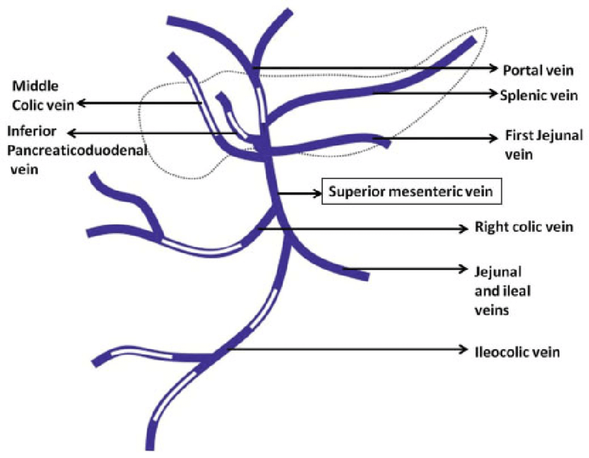
portal vein schematic

Parts:

  1. Supraduodenal
  2. Retroduodenal
  3. Infraduodenal

Tributaries:

  1. Splenic vein
  2. Superior mesenteric vein
  3. Cystic vein
  4. Paraumbilical vein
  5. Right and Left gastric vein
  6. Superior pancreaticoduodenal vein

Points to remember:

Tributaries of splenic vein:

  • Veins corresponding to the branches of splenic artery
  • Inferior mesenteric vein

Tributaries of inferior mesenteric vein:

  • Veins corresponding to the branches of inferior mesenteric artery

Tributaries of superior mesenteric vein:

  • Veins corresponding to the branches of superior mesenteric artery
  • Inferior pancreaticoduodenal vein
  • Right gastroepiploic vein

Sites of Portocaval Anastomoses

SitePortal veinSystemic veinPortal hypertension
Abdominal esophagusEsophageal tributaries of left gastric vein Esophageal tributaries of accessory hemiazygos veinEsophageal varices
UmbilicusParaumbilical veinSuperficial epigastric veinCaput medusae
Anal canalSuperior rectal veinMiddle and Inferior rectal veinHemorrhoids
Bare area of liverHepatic venulesPhrenic and intercostal veins
Posterior abdominal wallTwigs of colic veinRetroperitoneal vein
  • Facebook
  • Twitter
PGMEE, MRCS, USMLE, MBBS, MD/MS AnatomyGastrointestinal system

Post navigation

Previous post
Next post

Related Posts

PGMEE, MRCS, USMLE, MBBS, MD/MS

Thyroid Tumors : Mnemonics

Jun 17, 2023Jun 17, 2023

Papillary carcinoma Follicular carcinoma Medullary carcinoma Mnemonic: MENACED Anaplastic carcinoma

Read More
PGMEE, MRCS, USMLE, MBBS, MD/MS

Physiology of Pulmonary Surfactant

Jun 16, 2020Jun 16, 2020

Origin Type II alveolar cells/pneumocytes (small but numerous) and Lamellar bodies (appear in Type II pneumocytes at around the 20th week of gestation) Type I alveolar cells (large and covers 95% alveoli) are squamous cells and involved in gas exchange process. Recycling 90% is reprocessed (through endocytosis); average time for…

Read More
PGMEE, MRCS, USMLE, MBBS, MD/MS

Blood Supply of Talus

Jun 9, 2020Jun 9, 2020

3 Main Arteries Extraosseous and intraosseous anastomoses formed by 3 main arteries: Posterior tibial artery ~ 17% contribution Anterior tibial (dorsalis pedis) artery ~ 36% contribution Perforating peroneal artery ~ 47% contribution Major Branches Tarsal sinus artery: Arises from – Dorsalis pedis artery (frequently) or Lateral tarsal artery (branch of…

Read More

Leave a Reply Cancel reply

Your email address will not be published. Required fields are marked *

This site uses Akismet to reduce spam. Learn how your comment data is processed.

Epomedicine. Portal Vein : Tributaries and Portocaval Anastomoses [Internet]. Epomedicine; 2020 Sep 28 [cited 2025 Dec 6]. Available from: https://epomedicine.com/medical-students/portal-vein-tributaries-and-portocaval-anastomoses/.

Pre-clinical (Basic Sciences)

Anatomy

Biochemistry

Community medicine (PSM)

Embryology

Microbiology

Pathology

Pharmacology

Physiology

Clinical Sciences

Anesthesia

Dermatology

Emergency medicine

Forensic

Internal medicine

Gynecology & Obstetrics

Oncology

Ophthalmology

Orthopedics

Otorhinolaryngology (ENT)

Pediatrics

Psychiatry

Radiology

Surgery

RSS Ask Epomedicine

  • What to study for Clinical examination in Orthopedics?
  • What is the mechanism of AVNRT?

Epomedicine weekly

  • About Epomedicine
  • Contact Us
  • Author Guidelines
  • Submit Article
  • Editorial Board
  • USMLE
  • MRCS
  • Thesis
©2025 Epomedicine . All rights reserved.