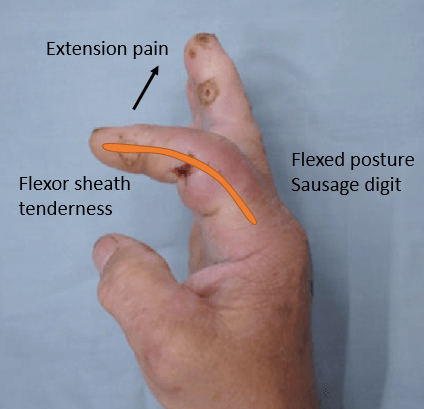
1. Exquisite tenderness over the course of the sheath, limited to the sheath Present in 64% cases Late sign of proximal extension of pyogenic tenosynovitis Most important sign as described by Kanavel 2. Flexion of the finger (‘hook’ sign) Present in 69% cases 3. Exquisite pain on extending the finger,…
Tag: Orthopedics
Section Editor: Dr. Sulabh Kumar Shrestha, MBBS, PGY1 Orthopedics
PGMEE, MRCS, USMLE, MBBS, MD/MS
Oestern and Tscherne Classification for Closed fractures
The classification system for closed fractures is based on the physiologic concept that the energy imparted to the bone (and the resultant fracture pattern) directly correlates with the energy transferred to the surrounding soft tissues. Grade Soft tissue injury Fracture Compartment C0 Absent or Negligible Simple (Spiral) Soft and/or Normal…
PGMEE, MRCS, USMLE, MBBS, MD/MS
Screw-home mechanism
Synonym: Knee-locking mechanism Definition: During the final degrees of knee extension (last 20-30 degrees), an obligatory lateral rotation of the tibia occurs, i.e. non-voluntary coupled movement of knee extension and external rotation. Mechanism: External rotation of tibia with reference to the femur or Internal rotation of femur with reference to…
PGMEE, MRCS, USMLE, MBBS, MD/MS
Foot muscles – Layers and Compartments
Deep to the plantar fascia, muscles of plantar foot exist in 4 different layers. Extensor digitorum brevis makes dorsal layer of foot and remaining 18 muscles and 4 tendons make the 4 layers of plantar aspect or sole of foot. In general: Layer 1 and 3: 3 muscles each that…
PGMEE, MRCS, USMLE, MBBS, MD/MS
Periprosthetic Joint Infection (PJI) Criteria and Management
The 2013 ICM criteria for defining Periprosthetic joint infection (PJI) can be remembered using the mnemonic below: Mnemonic: 1 tract or 2 bact. and 3 of ABCDEF 1 sinus tract communication with the joint OR 2 positive periprosthetic cultures with phenotypically identical bacteria AND 3 of the following six minor…
PGMEE, MRCS, USMLE, MBBS, MD/MS
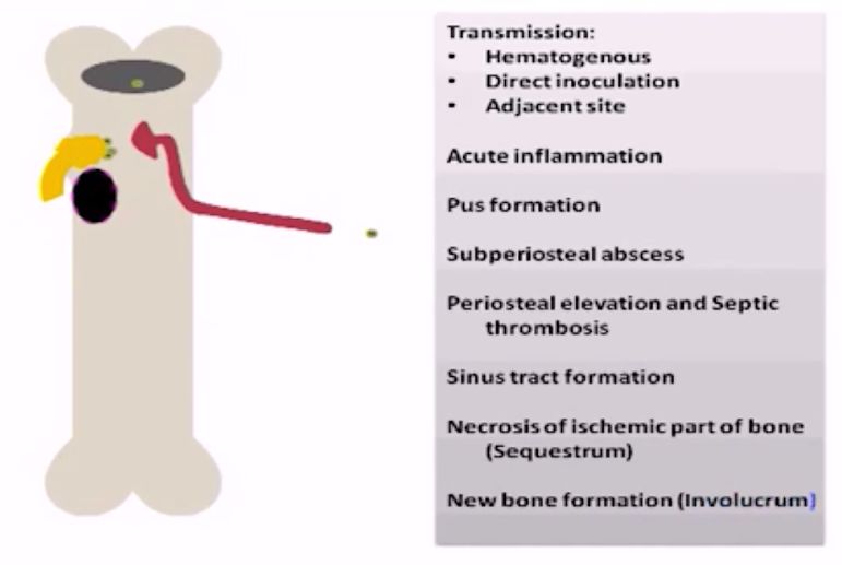
Pathophysiology of Osteomyelitis
Osteomyelitis is defined as an inflammation of the bone (single portion or numerous regions, like marrow, cortex, periosteum and surrounding soft tissue) caused by an infecting organism (usually monomicrobial but polymicrobial can occur, especially in diabetic foot). Definitions, Criteria and Classifications of different types of Osteomyelitis have been discussed in…
PGMEE, MRCS, USMLE, MBBS, MD/MS
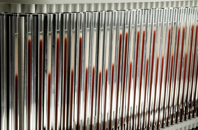
ESR and CRP in Musculoskeletal infection
ESR (Erythrocyte Sedimentation Rate) Usually elevated within 48-72 hours of the infection onset (less reliable in the first 48 hours of infection) Continues to rise for 3-5 days after institution of successful therapy and continuing rise beyond 4th-5th day of treatment can be an indication of treatment failure (not good…
Emergency Medicine

Polytrauma Assessment and Management (ATLS) : Mnemonics
SIEVE triage system Mnemonic: ABC-30-2 Can Do Approach to assessment and initial management Mnemonic: ARM Adjuncts to Primary Survey Reflects the adequacy of resuscitation. Mnemonic: PEA COVER Primary Survey Follow the look, listen, feel approach – Mnemonic: ABCDE 1. Airway and C-spine protection: 2. Breathing: 3. Circulation and control of…