3 Main Arteries
Extraosseous and intraosseous anastomoses formed by 3 main arteries:
- Posterior tibial artery ~ 17% contribution
- Anterior tibial (dorsalis pedis) artery ~ 36% contribution
- Perforating peroneal artery ~ 47% contribution
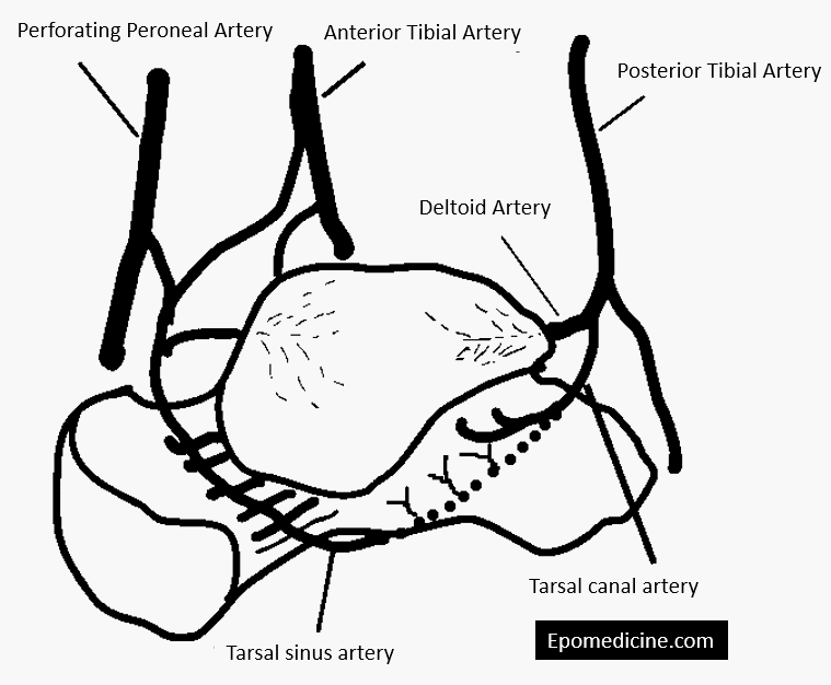
Major Branches
Tarsal sinus artery: Arises from –
- Dorsalis pedis artery (frequently) or
- Lateral tarsal artery (branch of dorsalis pedis artery) or
- Perforating peroneal artery or
- Anastomosis between Lateral tarsal artery and Perforating peroneal artery
Tarsal canal artery: Arsies from Posterior tibial artery
Deltoid artery: Arises from tarsal canal artery or posterior tibial artery
Superomedial direct branches: Arises from Dorsalis pedis artery
Posterior direct branches: Arises from the Posterior tibial artery
Major Anastomoses
1. Tarsal canal: Tarsal sinus artery + Tarsal canal artery = Major arterial axis of talus (arterial line is located posterior to the talar neck level)
2. Large periosteal network (continues into tarsal sinus and ends on the lateral side of body around lateral malleolus): Deltoid artery + Dorsalis pedis (superior neck branches)
3. Posterior tubercle: Peroneal artery + Posterior direct branches of posterior tibial artery
Sites of vascular access
More than half (almost two-third) of the talar surface is covered with articular cartilage and the blood supply of talus enters at 5 non-articular sites:
- Neck of talus
- Superior surface: Anterior tibial artery branches
- Inferior surface: Tarsal canal anastomosis between tarsal sinus and tarsal canal artery
- Sinus tarsi
- Medial side of body: Deltoid artery
- Posterior tubercle: Anastomosis around posterior tubercle
Intraosseous territory
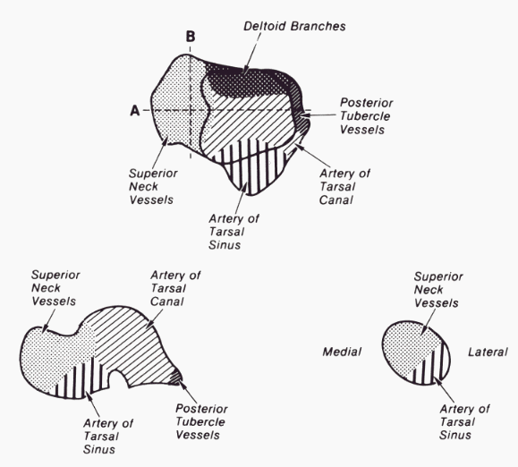
Head of talus:
- Supero-medially (2/3rd): from branches of dorsalis pedis artery
- Infero-laterally (1/3 rd): from tarsal sinus artery and it’s anastomosis or direct branches of lateral tarsal artery
Body of talus:
- Lateral 1/3: tarsal sinus artery
- Middle 1/3: anastomosis of tarsal sinus and tarsal canal artery
- Medial 1/3: deltoid artery
AVN in Tarsal neck fractures
Except for perfusion from the direct posterior branches and delotid branches, most of the talar body receives it’s blood in a retrograde fashion (distal to proximal) through anastomosis around the talar neck. Due to this reason, talar body is susceptible to avascular necrosis (AVN) in talar fractures. Non displaced fractures of talar neck disrupt intraosseous branches of tarsal sinus and tarsal canal, but the major vascular sling remains intact. With displaced fractures of talar neck, branches from dorsalis pedis artery to talar neck, tarsal canal artery & tarsal sinus artery (major arterial supply to talus) which are all connected through a sling of vessels within the tarsal sinus are vulnerable to rupture.
References:
1. Sarrafian’s Anatomy of the Foot and Ankle: Descriptive, Topographic, Functional By Armen S Kelikian
2. Mulfinger, G. L., & Trueta, J. (1970). THE BLOOD SUPPLY OF THE TALUS. The Journal of Bone and Joint Surgery. British Volume, 52-B(1), 160–167. doi:10.1302/0301-620x.52b1.160
3. Skeletal Trauma: Basic Science, Management, and Reconstruction, Volume 1 By Bruce D. Browner
4. Campbell’s Operative Orthopaedics, 13th Edition By S. Terry Canale, James H. Beaty