Skip to content
Epomedicine

Mnemonics, Simplified Concepts & Thoughts

Epomedicine

Mnemonics, Simplified Concepts & Thoughts

Singh Index for Osteoporosis – Simplified

Dr. Sulabh Kumar Shrestha, MS Orthopedics, Aug 8, 2021Aug 8, 2021

Pattern of bone loss in Osteoporosis

  1. Trabecular bone responds faster to metabolic changes due to greater surface are compared to cortical bone.
  2. Non-weight bearing (secondary trabeculae) are lost first. Weight bearing (primary trabeculae) may appear prominent due to loss of other trabeculae and may also become thickened due to compensatory mechanism or callus from microfractures.
  3. Later, even primary weight-bearing trabeculae are lost, resulting in translucent appearance of bone.
  4. In bones that sustain vertical loads, such as tibial and femoral metaphyses and vertebral bodies, resistance to lateral bowing and fractures is provided by a horizontal trabecular cross-bracing system that helps support the vertical elements. 

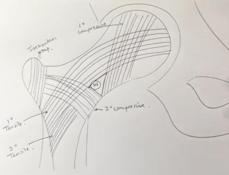
Trabeculae in Proximal Femur

5 groups based on orientation and function:

  1. Primary compressive group
  2. Secondary compressive group
  3. Greater trochanteric group
  4. Primary tensile group
  5. Secondary tensile group
femur trabeculae
Source: Quen Tang’s Youtube video on Trabecular Pattern

How do you remember where these trabeculae are ?

I use these general rules:

  1. Primary trabeculae start in head of femur as they are weight-bearing trabeculae.
  2. Secondary trabeculae lie inferior to the primary trabeculae.
  3. Compressive trabeculae go towards compression side of the proximal femur, i.e. medial side with lesser trochanter.
  4. Tensile trabeculae go towards tension side of the proximal femur, i.e. lateral side with greater trochanter.
  5. Greater trochanteric group is confined within greater trochanter.

Ward triangle refers to a radiolucent area between principal compressive, secondary compressive and primary tensile trabeculae in the neck of femur. This is the area having lowest BMD in femoral neck.
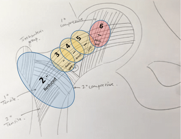
Singh’s Index

Based on the loss of trabecular pattern in proximal femur, Singh index is used to assist fracture prediction. As described by Singh index, trabecular bone loss in osteoporosis initiates in the trochanter, then evolves to the breakage of tensile trabeculae, and ultimately reaches the principal compressive trabeculae.

Grade 6 – All normal trabeculae groups visible

Grade 5 – Reduction/Loss of trochanteric and secondary trabeculae leading to: prominence of primary groups and enlargement of Ward’s triangle

Grade 4 – Reduced but continuous primary tensile group

Grade 3 – Discontinuous primary tensile group

Grade 2 – Loss of primary tensile group as well (Only primary compressive group visible)

Grade 1 – Reduction in primary compressive group

singhs index

However, Singh’s index has been found to poorly correlate with the BMD measured using DEXA or quantitative CT scan.

dr. sulabh kumar shrestha
Dr. Sulabh Kumar Shrestha, MS Orthopedics

He is the section editor of Orthopedics in Epomedicine. He searches for and share simpler ways to make complicated medical topics simple. He also loves writing poetry, listening and playing music. He is currently pursuing Fellowship in Hip, Pelvi-acetabulum and Arthroplasty at B&B Hospital.

18 shares
  • Facebook16
  • Twitter
PGMEE, MRCS, USMLE, MBBS, MD/MS OrthopedicsRadiology

Post navigation

Previous post
Next post

Related Posts

PGMEE, MRCS, USMLE, MBBS, MD/MS

Pedicle Screw Insertion Simplified

Oct 30, 2021Oct 30, 2021

Anatomy of Vertebral Pedicle Width (narrowest transverse diameter): Narrowest at T4-T5 (4-5 mm) Above and below this level, the width gradually increases to almost double at T1 and T11 (8 mm) Narrowest for lumbar at L2 (two for tiny; 2 X 3 = 6 mm) Increases gradually to L5 (5…

Read More
PGMEE, MRCS, USMLE, MBBS, MD/MS parotidectomy steps

Simplified Approach to Parotid Enlargement

Aug 7, 2015Aug 17, 2015

Diagnosis and management of a case of parotid enlargement requires systematic approach. Here, I have tried to present a simplified approach to parotid enlargement. A) DIFFERENTIAL DIAGNOSES OF PAROTID ENLARGEMENT: 1. Infectious: Child: Mumps, Coxsackie Adult: S.aureus 2. Non-infectious benign: Epithelial: Pleomorphic adenoma, Warthin tumor, Oncocytoma, Monomorphic adenoma Non-epithelial: Lymph…

Read More
PGMEE, MRCS, USMLE, MBBS, MD/MS choosing hypoglycemics

Approach to Uncomplicated Diabetes Mellitus : Simplified

Feb 19, 2016Jun 12, 2016

A) GLUCOSE LEVEL 1. Post-prandial: 140-200 mg/dl: Impaired glucose tolerance (Pre-diabetes) Diabetes prevented with: weight loss, exercise, metformin (in high risk) ≥200 mg/dl (+ Clinical symptoms): Diabetes confirmed 2. Fasting: 100-126 mg/dl: Impaired fasting glucose (Pre-diabetes) ≥126 mg/dl (+ Clinical symptoms): Diabetes confirmed

Read More

Comments (2)

  1. Dr vishwanath says:
    Jan 18, 2022 at 5:13 am

    What is the name of the person in Singh sintex ? Who is Singh ?

    Reply
    1. Epomedicine says:
      Jan 20, 2022 at 12:45 pm

      Dr. Manmohan Singh (Orthopedician) seems to be eponymous researcher behind the Singh’s Index.

      Singh M, Nagrath AR, Maini PS. Changes in trabecular pattern of the upper end of the femur as an index of osteoporosis. J Bone Joint Surg Am. 1970 Apr;52(3):457-67. PMID: 5425640.

      Reply

Leave a Reply to Dr vishwanath Cancel reply

Your email address will not be published. Required fields are marked *

This site uses Akismet to reduce spam. Learn how your comment data is processed.

Shrestha SK. Singh Index for Osteoporosis – Simplified [Internet]. Epomedicine; 2021 Aug 8 [cited 2025 Dec 19]. Available from: https://epomedicine.com/medical-students/singh-index-osteoporosis-simplified/.

Pre-clinical (Basic Sciences)

Anatomy

Biochemistry

Community medicine (PSM)

Embryology

Microbiology

Pathology

Pharmacology

Physiology

Clinical Sciences

Anesthesia

Dermatology

Emergency medicine

Forensic

Internal medicine

Gynecology & Obstetrics

Oncology

Ophthalmology

Orthopedics

Otorhinolaryngology (ENT)

Pediatrics

Psychiatry

Radiology

Surgery

RSS Ask Epomedicine

  • What to study for Clinical examination in Orthopedics?
  • What is the mechanism of AVNRT?

Epomedicine weekly

  • About Epomedicine
  • Contact Us
  • Author Guidelines
  • Submit Article
  • Editorial Board
  • USMLE
  • MRCS
  • Thesis
©2025 Epomedicine . All rights reserved.