Understanding of the Boutonniere and Swan neck deformity requires clear concept of the finger extensor apparatus, which has been discussed here.
The extrinsic extensor tendons trifurcate giving a central slip which attaches to the middle phalanx base and two lateral slips which join with lateral band (formed by intrinsic muscles – interossei +/- lumbricals) to form conjoined lateral tendon. The triangular ligament (of stack) connects the conjoined lateral band over the proximal phalanx. Transverse retinacular ligament connect the lateral bands to PIP joint volar plate. The conjoined tendons on either side converge at base of distal phalanx as terminal extensor tendon. The lateral bands lie volar to the axis of rotation of MCP joint and dorsal to the axis of rotation of interphalangeal joints, hence functions as MCP joint flexor and interphalangeal joint extensor. Oblique retinacular ligament (ORL) arises from volar plate of PIP joint and flexor tendon sheath and insert onto dorsal base of distal phalanx and interlinks the IP joint motion.
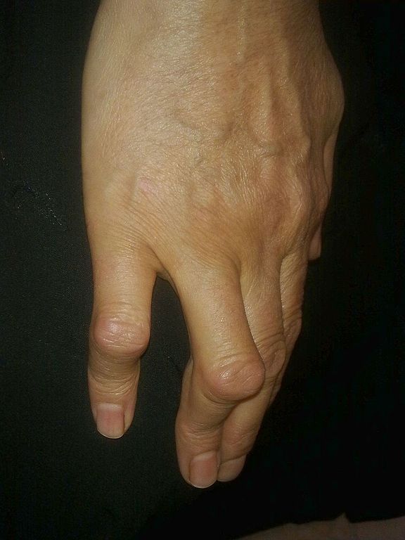
Boutonniere deformity
Flexion of PIP joint and hyperextension of DIP joint
1. Central slip injury:
- Loss of extrinsic extensor mechanism from EDC at PIP joint
- Active extension of PIP joint possible due to the action of conjoined lateral tendon if triangular ligament is intact
2. Triangular ligament disruption (acute injury or chronic attenuation) and volar subluxation of lateral band (due to pull of transverse retinacular ligament):
- The lateral band goes volar to the center of rotation of PIP joint
- Active extension of PIP joint is not possible (intrinsic mechanism is also lost)
- If passively extended, PIP joint can be held in position because on extension, the lateral band reverts back to their original position
- Unopposed pull of interossei and lumbricals cause DIP extension via terminal tendon
3. Oblique retinacular ligament contracture: Further increases DIP extension deformity
4. Shortening of collateral ligaments and volar plate of PIP joint: Permanent contracture and fibrosis of IP joint
Elson test for Central slip injury
Bend PIP 90° over edge of a table and extend middle phalanx against resistance. In the presence of central slip injury, there will be weak PIP extension and the DIP will go rigid due to lateral band activation. In the absence of central slip injury, lateral band remains inactivated and DIP joint remains floppy.
Hence, the management of Boutonniere deformity includes holding PIP joint in extension with splint, primary central band repair, terminal tendon tenotomy (modified Fowler or Dolphin), lateral band relocation, secondary tendon reconstruction, triangular ligament reconstruction and PIP arthrodesis.
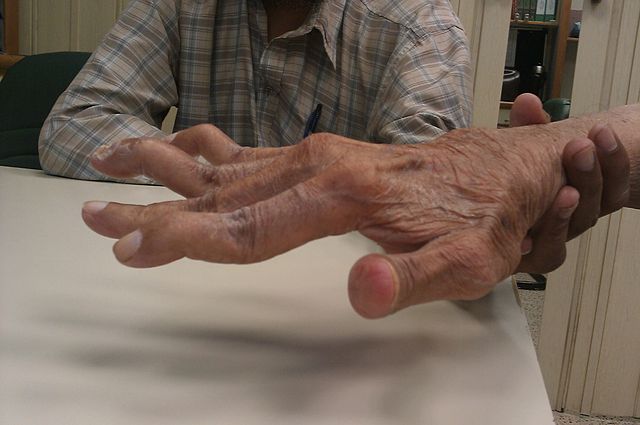
Swan neck deformity
Flexion of DIP joint and hyperextension of PIP joint
1. Secondary DIP flexion as a result of primary PIP hyperextension: PIP hyperextension can be –
a. Extrinsic type: Tightening of central slip due to excessive traction on extrinsic extensor tendon (EDC) due to muscle fibrosis, spasm, tendon adhesion or flexion deformity of wrist or MCP (elongates the course of extensor tendon)
b. Intrinsic type: Hyperactivity of intrinsic muscle (ischemic contracture, spasticity, intrinsic tendon adhesion, volar subluxation of MCP joint) overpowers the opposing action of extrinsic flexors
c. Articular type: Weakness of collateral ligaments, joint capsule, volar plate, FDS tendon, transverse retinacular ligament
PIP hyperextension relaxes the normal tension on lateral band leading to dorsal and central movement. With relaxation, lateral band loses the ability to extended the DIP joint. This leads to dropping of DIP into flexion.
PIP hyperextension also tensions FDP, leading to flexion of DIP joint.
Another contributing factor is the loss of the mechanical advantage of the oblique retinacular ligament in extending the DIP.
2. Primary DIP flexion deformity (mallet finger): Disruption or stretching of the terminal extensor tendon or the lateral band leading to flexion deformity of DIP –
All the power of extrinsic tendon (EDC) is concentrated on central slip. Over time, the structures of the PIP joint are weakened and the PIP joint is forced into hyperextension.
Bunnell test for Intrinsic tightness
Intrinsic tightness is demonstrated by comparing passive PIP flexion with MCP held in extension and flexed. With MCP extended, intrinsics are put on stretch and if they are tight, PIP joint will have limited flexion. If PIP joint flexion is restricted when MP joint is held in extension but not when MCP joint is held in flexion, patient has positive intrinsic tightness test. If PIP cannot be flexed even with MCP joint in flexion, then it is due to joint capsule tightness or contracture.
Hence, the management of swan neck deformity includes double ring splint to prevent PIP hyperextension, volar plate advancement for volar plate laxity, FDS tenodesis for FDS rupture, spiral oblique retinacular reconstruction and central slip tenotomy (Fowler).

He is the section editor of Orthopedics in Epomedicine. He searches for and share simpler ways to make complicated medical topics simple. He also loves writing poetry, listening and playing music. He is currently pursuing Fellowship in Hip, Pelvi-acetabulum and Arthroplasty at B&B Hospital.