Skip to content
Epomedicine

Mnemonics, Simplified Concepts & Thoughts

Epomedicine

Mnemonics, Simplified Concepts & Thoughts

Surface Anatomy of Pleura and Lung

Epomedicine, May 19, 2024

Surface Anatomy of Pleura

Mnemonic: 2, 4, 6, 8, 10, 12 rule

1. Starts about 2 cm above the midpoint of medial 1/3 of clavicle.

2. Meet in the midline at rib 2.

3. Left side reaches sternal line at rib 4 (to make room for heart).

4. Right side reaches sternal line at rib 6.

5. Both cross rib 8 in midclavicular line.

6. Both cross rib 10 in mid-axillary line.

7. Both cross mid-scapular line (posteriorly) in rib 12.

lung and pleura surface markings
“This image is taken from Page 17 of Surgery of the lung and pleura” by Medical Heritage Library, Inc. is licensed under CC BY-NC-SA 2.0.

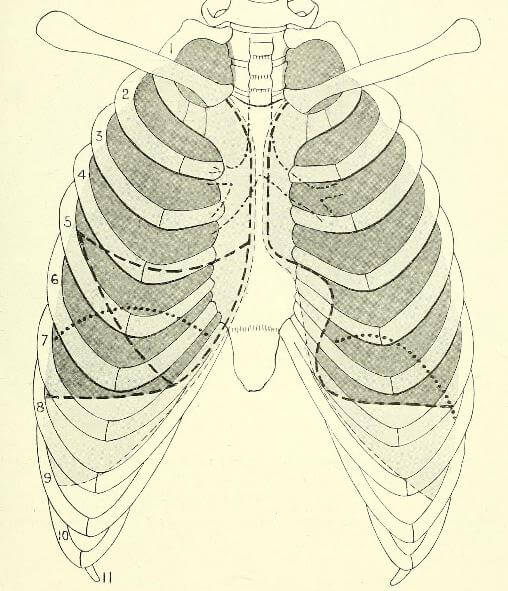
Surface Anatomy of Lungs

1. Apex, Anterior border and Posterior border of lungs: Correspond to the lines of pleura but are slightly away from median plane

2. Inferior margin:

Below rib 6, the lungs extend to two rib spaces less than pleura (i.e. opposite to rib 6 mid-clavicular line, rib 8 mid-axillary line and rib 10 posteriorly). The parietal pleura extends a further two rib spaces inferiorly than the inferior lung edge to allow space for lung expansion.

LevelParietal pleuraLungs
Midclavicular line8th rib6th rib
Midaxillary line10th rib8th rib
Midscapular line12th rib10th rib

3. Oblique fissure: T3 to 6th costal cartilage

4. Transverse fissure (right lung only): Right 4th costal cartilage (sternal edge) to Oblique fissure

  • Facebook
  • Twitter
PGMEE, MRCS, USMLE, MBBS, MD/MS AnatomyRespiratory system

Post navigation

Previous post
Next post

Related Posts

PGMEE, MRCS, USMLE, MBBS, MD/MS Appendicular-skeleton

Summary of Clinical Tests and Signs in Orthopedics

Jun 5, 2014

Adson’s test: for thoracic outlet syndrome Allen’s test: for testing patency of radial and ulnar arteries Alli’s test: for DDH Anvil test: for testing tenderness of the spine Ape thumb: for median nerve injury Apley’s grinding test: for meniscus injury Apprehension test: for recurrent dislocation of the shoulder Barlow’s test:…

Read More
PGMEE, MRCS, USMLE, MBBS, MD/MS pleural effusion xray

Chest X-ray – Pleural Effusion

Jun 21, 2015

Pleura is a mesothelial lined sac that envelopes the lungs and comprises of 2 membranous walls i.e. visceral pleura and parietal pleura that encloses pleural space filled with pleural fluid. Pleural space contains about 0.3 ml/kg body weight of pleural fluid. The pleura is not visible on a normal CXR…

Read More
PGMEE, MRCS, USMLE, MBBS, MD/MS

Male Reproductive Anatomy : Mnemonics

May 13, 2024May 18, 2024

Penile Layers Mnemonic: ABCD (from inside to outside) 1. Albuginea (tunica albuginea): Envelopes 3 corporal bodies (2 cavernosa and 1 spongiosum) 2. Buck’s fascia: Separates corpora cavernosa from corpus spongiosum; separates superficial and deep dorsal veins of the penis 3. Colle’s fascia: Continuous with Scarpa’s fascia of abdomen and Dartos…

Read More

Leave a Reply Cancel reply

Your email address will not be published. Required fields are marked *

This site uses Akismet to reduce spam. Learn how your comment data is processed.

Pre-clinical (Basic Sciences)

Anatomy

Biochemistry

Community medicine (PSM)

Embryology

Microbiology

Pathology

Pharmacology

Physiology

Clinical Sciences

Anesthesia

Dermatology

Emergency medicine

Forensic

Internal medicine

Gynecology & Obstetrics

Oncology

Ophthalmology

Orthopedics

Otorhinolaryngology (ENT)

Pediatrics

Psychiatry

Radiology

Surgery

RSS Ask Epomedicine

  • What to study for Clinical examination in Orthopedics?
  • What is the mechanism of AVNRT?

Epomedicine weekly

  • About Epomedicine
  • Contact Us
  • Author Guidelines
  • Submit Article
  • Editorial Board
  • USMLE
  • MRCS
  • Thesis
©2026 Epomedicine | WordPress Theme by SuperbThemes