Skip to content
Epomedicine

Mnemonics, Simplified Concepts & Thoughts

Epomedicine

Mnemonics, Simplified Concepts & Thoughts

pons cross-section

How to Draw Pons Cross-section ?

Dr. Sulabh Kumar Shrestha, MS Orthopedics, Jul 19, 2016Sep 1, 2020

Pons in latin, refers to a “bridge”. Pons varolli is a part of brain-stem, that links thalamus with medulla oblongata. The cross-section of pons is similar to the midbrain as described earlier but few things must be kept in mind:

  1. The orientation of lemnisci in midbrain is more or less vertical, but in pons it is horizontal.
  2. Cranial nerve III and IV arises from midbrain and mainly Cranial nerve V, VI, VIII and VIII arises from pons.
  3. Cerebral aqueduct lies in midbrain and 4th ventricle lies in pons.

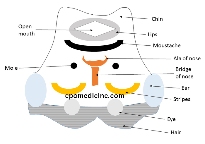
Earlier, we imagined transverse section of the midbrain as an inverted striped face of red-eyed demon. Similary, we will use the analogy of an “inverted face of a human” to draw the cross-section of pons.
pons mnemonic

Now, let’s label the structures shown above:

  1. Hair = Transverse pontine fibers
  2. Eye = Corticospinal and Corticonuclear tracts
  3. Ear = Middle cerebellar peduncle
  4. Stripes = Lemnisci
    • Medially: Medial lemniscus
    • Middle: Trigeminal lemniscus medially and Spinal lemniscus laterally
    • Lateral: Lateral lemniscus
  5. Bridge of nose = Raphe nucleii
  6. Ala of nose = Medial Longitudinal Fasciculus
  7. Mole = Facial nerve motor nucleus (In caudal pons)
  8. Moustache = Cranial nerve nucleii
    • Medial most = CN VI or Abducens nerve (In caudal pons)
    • Middle = CN V or Trigeminal nerve – motor and sensory (In rostral pons)
    • Lateral most = CN VIII – Superior vestibular nucleus (In caudal pons)
  9. Lips = Periventricular gray
    • Contains locus coeruleus
  10. Open mouth = 4th ventricle
  11. Chin = Superior cerebellar peduncle

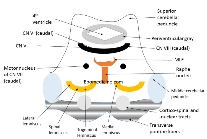
Now, let’s look at the real picture:
pons cross-section

You can compare with your own face to learn the pons. Wasn’t that easy? Leave comments below 🙂 .

dr. sulabh kumar shrestha
Dr. Sulabh Kumar Shrestha, MS Orthopedics

He is the section editor of Orthopedics in Epomedicine. He searches for and share simpler ways to make complicated medical topics simple. He also loves writing poetry, listening and playing music. He is currently pursuing Fellowship in Hip, Pelvi-acetabulum and Arthroplasty at B&B Hospital.

8 shares
  • Facebook6
  • Twitter
PGMEE, MRCS, USMLE, MBBS, MD/MS AnatomyNervous system

Post navigation

Previous post
Next post

Related Posts

PGMEE, MRCS, USMLE, MBBS, MD/MS

Mnemonic Approach to Elbow Xray – FOOL

Oct 12, 2019Jun 14, 2022

Mnemonic: FOOLa. Fat padsb. Overt findings and outlinesc. Ossification centersd. Lines Fat pads The elbow joint has anterior and posterior fat pads which are extra-synovial but within the articular capsule. These are present as a radiolucent (black) line (compared to bone and surrounding soft tissues) in lateral X-rays. Normal finding:…

Read More
PGMEE, MRCS, USMLE, MBBS, MD/MS bartter-gitelmann-liddle

Bartter, Gitelmann and Liddle syndrome

Jun 6, 2019Jun 6, 2019

The order of the nephron defects in Bartter, Gitelmann and Liddle syndrome follows alphabetical order. Bartter: Thick ascending LOHGitelmann: Distal tubuleLiddle: Collecting duct a. Bartter syndrome: Thick ascending Loop of Henle (LOH) Defect in Na-2K-Cl transporter (like loop diuretics) NaCl wasting, Hypercalciuria and mild hypomagnesemia Mnemonic: Loop diuretics lose calcium…

Read More
PGMEE, MRCS, USMLE, MBBS, MD/MS omental cake

Omental Cake

Jul 11, 2016

Definition of Omental Caking Thickening of the omentum resulting from localized or diffuse infiltration of omental fat by soft tissue density mass is referred as “omental caking”. It is a radiological sign, which is often identified in CT scan. Involved Anatomical Structure in Omental Caking Greater Omentum – an extension of…

Read More

Comments (6)

  1. Aishwarya says:
    Jun 12, 2018 at 2:11 pm

    Helpful!! Thanks a lot

    Reply
  2. Ana says:
    May 13, 2020 at 7:46 pm

    This is so helpful and creative! I loved the midbrain red-eyed demon too!

    Reply
  3. THARUN TEJ says:
    Sep 1, 2020 at 1:44 am

    hii sir, could u please check whether the location of superior vestibular nucleus is at caudal level or rostral level,
    i confused a lot regarding this , and i think its in causal part of pons , pls verify this sir

    Reply
    1. Dr. Sulabh Kumar Shrestha, MBBS, PGY1 Orthopedics says:
      Sep 1, 2020 at 2:28 am

      Hello Tharun, thank you for the correction. The four vestibular nuclei located in the rostral medulla and caudal pons. Superior and lateral vestibular nuclei are loacted in the caudal pons. Superior vestibular nucleus is located entirely within caudal pons while the lateral vestibular nucleus extends from the caudal pons to rostral medulla.

      Reference: A Textbook of Neuroanatomy By Maria A. Patestas, Leslie P. Gartner

      Reply
  4. Mahmud Musa says:
    Nov 24, 2023 at 6:31 am

    Thank you sir, love from Nigeria!

    Reply
    1. Raphael says:
      May 1, 2024 at 10:14 pm

      This website has been so helpful.
      ♥️ From a fellow Nigerian.

      Reply

Leave a Reply Cancel reply

Your email address will not be published. Required fields are marked *

This site uses Akismet to reduce spam. Learn how your comment data is processed.

Shrestha SK. How to Draw Pons Cross-section ? [Internet]. Epomedicine; 2016 Jul 19 [cited 2026 Aug 24]. Available from: https://epomedicine.com/medical-students/draw-pons-cross-section/.

Pre-clinical (Basic Sciences)

Anatomy

Biochemistry

Community medicine (PSM)

Embryology

Microbiology

Pathology

Pharmacology

Physiology

Clinical Sciences

Anesthesia

Dermatology

Emergency medicine

Forensic

Internal medicine

Gynecology & Obstetrics

Oncology

Ophthalmology

Orthopedics

Otorhinolaryngology (ENT)

Pediatrics

Psychiatry

Radiology

Surgery

RSS feed: Ask Epomedicine Ask Epomedicine

  • What to study for Clinical examination in Orthopedics?
  • What is the mechanism of AVNRT?

Epomedicine weekly

  • About Epomedicine
  • Contact Us
  • Author Guidelines
  • Submit Article
  • Editorial Board
  • USMLE
  • MRCS
  • Thesis
©2026 Epomedicine | WordPress Theme by SuperbThemes