Skip to content
Epomedicine

Mnemonics, Simplified Concepts & Thoughts

Epomedicine

Mnemonics, Simplified Concepts & Thoughts

Circle of willis mnemonic

Circle of Willis – Mnemonic and Drawing

Dr. Sulabh Kumar Shrestha, MS Orthopedics, Aug 1, 2016May 18, 2024

Circle of Willis is an important arterial communication that supplies the forebrain (telencephalon, diencephalon and optic vesicle) and often frequently tested in the exams. Circle of Willis receives blood from:

  1. Vertebrobasilar system: Basilar artery which gives off Posterio Cerebral Arteries (PCA) and Posterior communicating arteries which are the branches of PCA
  2. Internal Carotid Artery System: Gives off other arteries of Circle of Willis

Here, we will learn a mnemonic to draw the circle of willis and intracranial course of Internal Carotid Artery (ICA).
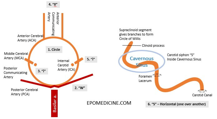
Circle of willis mnemonic

Step-wise instructions for drawing the circle of willis:

1. Draw a Circle – Circle of Willis is a circle of arteries.

Now write the “Willis” around this circle:

2. Write a large “W” at the inferior of circle – this represents Posterior Cerebral Arteries (PCA) which arises from a single basilar artery.

3. Write a horizontal “i” at the sides of the circle – this represents Middle Cerebral Artery (MCA) outside the circle and Internal Cerebral Artery (ICA) inside the circle.

4. Write “l l” at the superior of the circle – this represents the Anterior Cerebral Arteries (ACA) along with the part of circle between the MCA-ICA and Anterior communicating artery.

5. Write a horizontal “i” at the sides of the circle – this represents Middle Cerebral Artery (MCA) outside the circle and Internal Cerebral Artery (ICA) inside the circle.

Now, the intra-cranial course of Internal Carotid Artery:

6. Write a horizontal “S” – starting from carotid canal and ending in foramen lacerum.

7. Write another horizontal “S” beginning from the end of previous “S” –

  • starting from foramen lacerum and then
  • forming “S” shaped carotid siphon within cavernous sinus and then
  • ending at the level of anterior clinoid process
  • supraclinoid segment of ICA gives of branches to form Circle of Willis
ArteryFunction/supply
Anterior cerebral (ACA)Frontal lobes, Superior-Medial parietal lobes
Anterior communicatingConnects ACA
Middle cerebral (MCA)Lateral cerebral hemispheres (mostly), Basal ganglia, Internal capsule
Posterior communicatingJoins PCA to terminal trifurcation of ICA
Posterior cerebral (PCA)Occipital lobes
Superior cerebellar (SCA)Superior cerebellum, Parts of midbrain
BasilarBrainstem
Anterior Inferior Cerebellar (AICA)Anterior inferior cerebellum
VertebralDeliver blood to circle of Willis
dr. sulabh kumar shrestha
Dr. Sulabh Kumar Shrestha, MS Orthopedics

He is the section editor of Orthopedics in Epomedicine. He searches for and share simpler ways to make complicated medical topics simple. He also loves writing poetry, listening and playing music. He is currently pursuing Fellowship in Hip, Pelvi-acetabulum and Arthroplasty at B&B Hospital.
8 shares
  • Facebook8
  • Twitter
PGMEE, MRCS, USMLE, MBBS, MD/MS AnatomyNervous system

Post navigation

Previous post
Next post

Related Posts

PGMEE, MRCS, USMLE, MBBS, MD/MS ulnar nerve course

Ulnar nerve Anatomy – Course and Innervation

Jun 7, 2018Jun 1, 2024

Origin: C(7), C8, T1 (medial cord of Brachial plexus) Course: Motor innervation: 1. Forearm: Flexor carpi ulnaris (weakness of ulnar deviation and flexion of wrist), Medial half of flexor digitorum profundus (branches near the elbow) 2. Hand: branches near wrist Sensory innervation: Palmar branch at forearm and Digital branch at wrist…

Read More
PGMEE, MRCS, USMLE, MBBS, MD/MS

JVP Mnemonics

Aug 4, 2019Aug 4, 2019

Jugular venous pulse (JVP) waveforms a – Atrial contraction (RA) c – Closure and Curving of tricuspid valve into RA x – atrial relaXation v – Venous filling of right atrium (RA) y – atrial emptYing Distinguishing JVP from Internal carotid pulsation Mnemonic: POLICE Palpation: Non-palpable Occlussion: Readily occludable Location:…

Read More
PGMEE, MRCS, USMLE, MBBS, MD/MS

Supports of Uterus : Mnemonic

May 25, 2023May 25, 2023

Primary Supports 1. Mechanical Support Mnemonic: 3 A 2. Muscular/Active Support Mnemonic: Muscle PULLs 3. Ligamentary Support Mnemonic: Ligaments hold uterus like CUP Cardinal ligament + Pubocervical ligament = Triradiate ligament Secondary supports (Peritoneal folds) Mnemonic: RUB

Read More

Leave a Reply Cancel reply

Your email address will not be published. Required fields are marked *

This site uses Akismet to reduce spam. Learn how your comment data is processed.

Shrestha SK. Circle of Willis – Mnemonic and Drawing [Internet]. Epomedicine; 2016 Aug 1 [cited 2026 Jul 22]. Available from: https://epomedicine.com/medical-students/circle-willis-mnemonic-drawing/.

Pre-clinical (Basic Sciences)

Anatomy

Biochemistry

Community medicine (PSM)

Embryology

Microbiology

Pathology

Pharmacology

Physiology

Clinical Sciences

Anesthesia

Dermatology

Emergency medicine

Forensic

Internal medicine

Gynecology & Obstetrics

Oncology

Ophthalmology

Orthopedics

Otorhinolaryngology (ENT)

Pediatrics

Psychiatry

Radiology

Surgery

RSS Ask Epomedicine

  • What to study for Clinical examination in Orthopedics?
  • What is the mechanism of AVNRT?

Epomedicine weekly

  • About Epomedicine
  • Contact Us
  • Author Guidelines
  • Submit Article
  • Editorial Board
  • USMLE
  • MRCS
  • Thesis
©2026 Epomedicine | WordPress Theme by SuperbThemes