We have already discussed earlier on the intracranial course of Internal Carotid Artery (ICA) and Circle of Willis formation with the help of a simple mnemonic.
General Concepts of Blood Supply of Brain and Spinal Cord
1. Spinal cord, Hind-brain and Mid-brain: Veterbro-basilar system
2. Forebrain: Circle of willis which comprises of –
- Vertebrobasilar system: Posterior Cerebral Arteries (PCA) and it’s branch Posterior communicating artery
- Internal carotid system: Other arteries of circle of willis including Anterior Cerebral Arteries (ACA), Middle Cerebral Arteries (MCA) and Posterior Cerebral Arteries (PCA)
Forebrain derivatives are Telencephalon, Diencephalon and Optic vesicle.
3. The major vessels have 2 branches:
- Leptomeningeal branches: supply superficial regions (cortical and subcortical regions)
- Perforating branches: supply deep structures like diencephalon, basal ganglia and internal capsule)
4. Segments of the arteries:
- Proximal segments (A1 of ACA and P1 of PCA) are proximal to the respective communicating arteries, i.e. Anterior communicating artery and Posterior communicating artery respectively.
- Proximal segment of MCA (M1) is proximal to the bifurcation of trifurcation of the artery.
- Distal segments (A3 of ACA, M4 of MCA and P4 of PCA) are the cortical segments that supply the cortex.
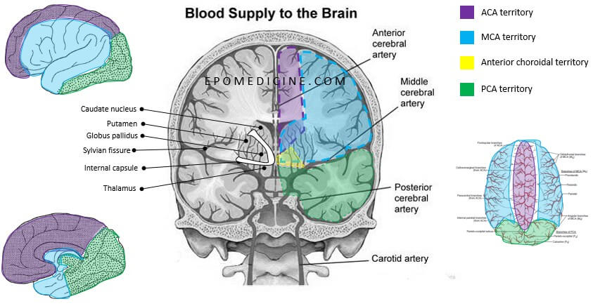
ICA enters through carotid canal to the cavernous sinus as shown in picture above.
5. Leptomeningeal territories of arteries in general:
- PCA: supplies posterior and inferior surfaces
- ACA: supplies antero-medial surface
- MCA: supplies lateral surface
Location of Circle of Willis
The arteries get interconnected around the interpeduncular fossa which is a rhomboid (diamond) shaped space in the ventral surface of the brain formed by the 2 Optic tracts anteriorly and 2 cerebral peduncles from pons posteriorly. It encloses tuber-cinerum, mammillary bodies, posterior perforated substance and the occulomotor neve from anterior to posterior.
Interpeduncular cistern or Basal cistern = Interpeduncular fossa + Cerebral peduncles + Circle of Willis
Importance of Circle of Willis
Collateral circulation
Anterior Cerebral Artery (ACA)
ACA runs in the interhemispheric fissure and arches anterior to the genu of corpus callosum and then along the cingulate sulcus to give off branches.
Remember: The course of ACA is more on medial and hence, have major supply on medial brain surface and also gives of medial striate branch as deep perforating branch.
Supplies:
- Medial surface of cerebral hemispheres
- Medial, infero-medial and supero-lateral parts of frontal lobe and medial parietal lobe
- Corpus callosum except splenium
- Caudate nucleus head, anteromedial and inferior basal ganglia, anterior limb and genu of inferior internal capsule
Middle Cerebral Artery (MCA)
MCA courses laterally on lateral sulcus or sylvian fissure (overlies the insula) and emerges out on the lateral surface of the brain. Suprasylvian branches supply lateral and inferior frontal lobe and anterior lateral parts of parietal lobe. Infrasylvain branches supply lateral temportal lobe including its anterior tip and the amygdala and posterior parietal.
Remember: The course of MCA is more on lateral and hence, have major supply on lateral brain surface and also gives lateral striate branch as deep perforating branch.
Supplies:
- Supplies lateral portion of basal ganglia.
- Superior surface of whole internal capsule
- Anterior temporal lobe and most of the lateral surface of the cerebral hemispheres
Posterior Cerebral Artery (PCA)
PCA courses backwards, beneath the splenium of corpus callosum, to the calcarine fissure and to the lateral occipital surface.
Remember: The course of PCA is backwards and hence, have major supply on the posterior brain surface. It gives thalamoperforating artery, thalamogeniculate artery and posterior chorodial artery as deep branches to supply the thalamus.
Supplies:
- Medial surface of parietal lobe
- Medial and inferior surface of temporal lobe including hippocampal formation
- Occipital lobe
- Splenium of corpus callosum
- Choroid plexus of ventricles
- Thalamus, hypothalamus and subthalamic nuclei
- Midbrain
Anterior Choroidal Artery
Branch of Internal Carotid Artery (ICA). It supplies:
- Posterior limb of inferior part of internal capsule
- Anterolateral thalamus
- Globus pallidus
- Posterior part of caudate nucleus and putamen
- Choroid plexus of anterior part of lateral ventricles
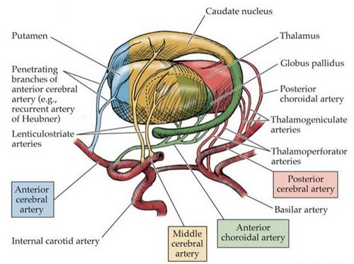
Deep Perforating Branches and Blood Supply of Basal Ganglia and Diencephalon
Circle of Willis Lesions
Berry Aneurysms
Commonest site is the branch point between anterior communicating artery and anterior cerebral artery (40%). Other common sites are the bifurcations/trifurcations of MCA (34%) and junction between posterior communicating artery and MCA (20%).
Branching points are the sites where arterial media are congenitally attenuated.
Remember the relations in interpeduncular fossa:
- Aneurysms in anterior communicating artery: compresses the optic tract (visual field defect – bitemporal lower quardantopia)
- Aneurysms in the posterior communicating artery: compresses the occulomotor nerve (CN III palsy – down and out eye)
Watershed Zones
Watershed zones are the border zones that receive dual blood supply from terminal or distal branches of the 2 large arteries. Strokes in these areas (watershed stroke or infarction) occurs during severe hypotension when the cerebral autoregulation fails and these border zones are starved of blood.
The somatotopic sensorimotor area between the middle and anterior cerebral arteries encodes the proximal arms and legs. So patients with watershed strokes often manifest with “man in a barrel syndrome,” meaning they have trouble lifting their arms and legs but their hands and feet are fine.
Anterior Cerebral Artery (ACA)
The sensorimotor homnculus for leg and foot are in the paracentral lobule in the medial aspect of brain. Hence, the lesion leads to contralateral leg-foot motor and sensory loss.
ACA also supplies the majority of anterior portion of corpus callosum, the damage of which leads to transcortical apraxia (a disconnect syndrome). There is no motor weakness, but the patient cannot execute a command to move their left arm.
- Wernicke’s area intact in left hemisphere: can understand the command
- Callosal lesion: disconnection of wernicke’s area from right primary motor cortex – cannot execute left arm movement on command.
- Connection between wernicke’s area and left primary motor cortex intact: can execute a command to move right arm
Middle Cerebral Artery (MCA)
MCA supplies the lateral convexity (parietal region) of the brain including:
- Broca’s and Wernicke’s speech areas
- Arcuate fasciculus or Superior longitudinal fasciculus
- Sensorimotor homonculus of remaining areas, i.e. face and arms
- Frontal eye field
- Splenium of corpus callosum
Lesion leads to:
- Contralateral face and arm paralysis and sensory loss
- Dominant hemisphere (Speech centers): Aphasia
- Non-dominant hemisphere (Supramarginal gyrus and angular gyrus – body and spatial awareness): Contralateral hemisensory neglect
Posterior Cerebral Artery (PCA)
Supplies occipital cortex, diencephalon and rostral midbrain.
Lesion leads to contralateral homonymous hemianopia with macular sparing and alexia without agraphia if the lesion is on left side (dominant hemisphere). Alexia results as the visual information from intact right (non-dominant) cortex is blocked by the lesion and cannot reach the language area. This is due to damage of splenium of corpus callosum.
Lateral Striate Artery
Branch of MCA which supplies the anterior limb of the internal capsule (supply superior portion of internal capsule including posterior limb which project corticospinal fibers) and ventral thalamus (relays sensory tracts).
- Isolated infarction of internal capsule: Pure motor stroke (contralateral hemiparesis)
- Isolated infarction of thalamus: Pure sensory stroke (contralateral sensory loss)
Other lesions
Previously, we have discussed about the various brodmann area’s location and functions. Damage to these areas have specific pathology.