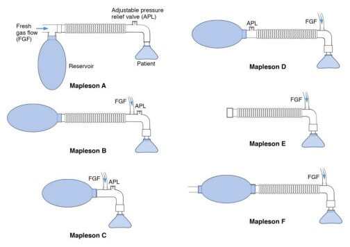
Anatomy of Mapleson Breathing Circuit Basically, a mapleson breathing circuit consists of following parts: 1. Face mask (towards patient end) 2. Reservoir bag (towards operator end) Accommodates fresh gas flow during expiration acting as a reservoir available for the following inspiration. Acts as a monitor of patient’s ventilatory pattern during…
Category: PGMEE, MRCS, USMLE, MBBS, MD/MS
Medical knowledge in bullet points with understandable language, simplified images and graspable mnemonics.
PGMEE, MRCS, USMLE, MBBS, MD/MS
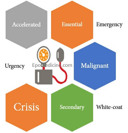
Terminologies of Hypertension
There are various terminologies used to describe hypertension which may overlap and are a source of confusion to the medical students and health professionals. Essential or Primary or Idiopathic hypertension Hypertension in which secondary causes have been excluded. Identifiable etiologic factors of essential hypertension: Obesity Insulin resistance High alcohol intake…
PGMEE, MRCS, USMLE, MBBS, MD/MS
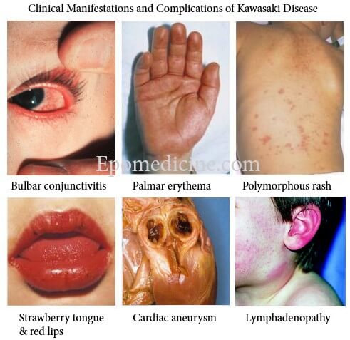
Kawasaki Disease – Diagnostic Criteria Mnemonic
The diagnostic criteria of Kawasaki Disease can be remembered using a mnemonic – “FEBRILE“. Fever: >5 days plus ≥4 of the following Enathem: Lips: Erythema, fissuring or crusting Oropharynx: Diffuse injection Tongue: Strawberry tongue Bulbar conjunctivitis: Bilateral, painless and non-exudative Rash: Polymorphous rash Internal organ involvement (not the part of criteria)…
PGMEE, MRCS, USMLE, MBBS, MD/MS
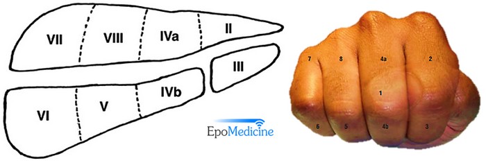
Liver Segments Explained with Mnemonic
Couniaud divided liver into 8 functional segments, each of which is supplied by it’s own portal triad (composed of a portal vein, hepatic artery and a bile duct). Hepatic veins divide the liver in saggital plane: 1. Middle hepatic vein: Divides the liver into right and left functional lobe. 2….
PGMEE, MRCS, USMLE, MBBS, MD/MS
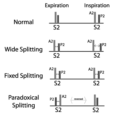
Abnormalities of First and Second Heart Sound
In the chapter of cardiac cycle, we have discussed the mechanism of production of heart sounds and their physiologic splitting. First Heart Sound (S1) Mechanism Closure of atrioventricular valves. It is best appreciated in mitral and tricuspid area of chest for respective components. Loud S1 Slamming a door from a…
PGMEE, MRCS, USMLE, MBBS, MD/MS
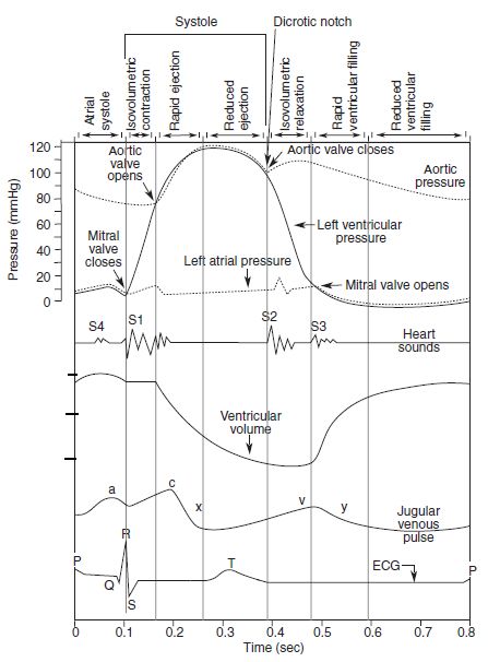
Cardiac Cycle – Summary and Wigger’s Diagram
Cardiac Cycle Opening and closing of valves When the valve opens, different compartments act as a single chamber (atrio-ventricle or aorto-ventricle). For a blood to flow, pressure in “giver” must be higher then that in “receiver”. Pressure difference opens or closes the valve: Role of atrial contraction in Ventricular filling…
PGMEE, MRCS, USMLE, MBBS, MD/MS
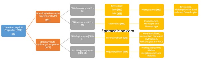
Concept of Acute Myeloid Leukemia (AML) FAB Classification
There is no need of mnemonics to remember the FAB classification of Acute Myeloid Leukemia (AML); just remember the process myeloid differentiation. A simple schematic diagram with few intermediate processes and stimulating factors eliminated will meet our purpose here. The cells belonging to the myeloid lineage are: Granulocytes: Neutrophils, Eosinophils…
PGMEE, MRCS, USMLE, MBBS, MD/MS
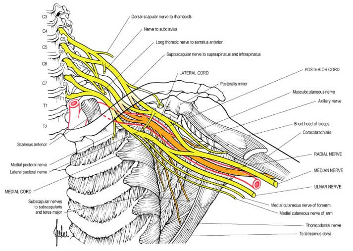
Brachial Plexus Simplified with Mnemonics
Components of Brachial Plexus Mnemonic: Randy Travis Drinks Cold Beer From proximal to distal, brachial plexus consists of: How are the roots formed? From the Ventral Rami of C5 to T1 spinal nerves. Extent and course: Intervertebral foramina to Transverse process to Interscalene triangle (bounded by anterior scalene and middle…