Synonyms: Connexus intertendinei, Intertendinous connections Plural: Juncturae tendinae Location: Intermetacarpal spaces in dorsum of hand between the extensor digitorum tendons Morphologic types: The usual pattern is that it gets thicker from radial to ulnar side. Type 1: Thin filamentous (square, rhomboidal or triangular) – present only in 2nd metacarpal space…
Category: PGMEE, MRCS, USMLE, MBBS, MD/MS
Medical knowledge in bullet points with understandable language, simplified images and graspable mnemonics.
PGMEE, MRCS, USMLE, MBBS, MD/MS
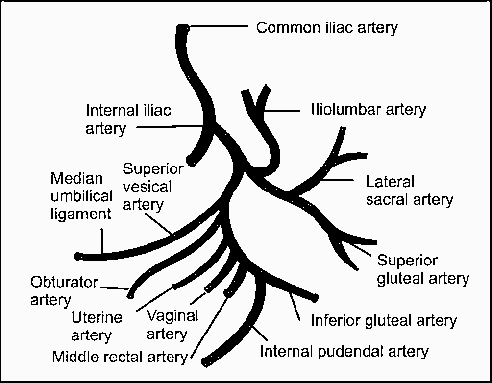
Internal Iliac Artery Anatomy : Simplified
Origin L5-S1 (common iliac artery bifurcation; anterior to SI joint) Course Extends down and posteriorly ~4 cm until superior margin of greater sciatic foramen and bifurcates into 2 trunks (in 60% cases) – Anterior trunk: continuation of the main artery towards ischial spine Posterior trunk: passes towards greater sciatic foramen…
PGMEE, MRCS, USMLE, MBBS, MD/MS
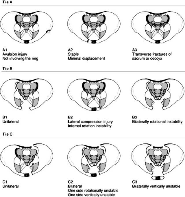
Pelvic Fracture Classification and Management : Simplified
Before proceeding to this topic, it would be wise to go through the topics listed below: Tile/AO Classification Tile classification divides pelvic fractures into three basic types according to stability based on the integrity of the posterior sacroiliac complex. Here is a mnemonic that can be used to remember tile…
PGMEE, MRCS, USMLE, MBBS, MD/MS
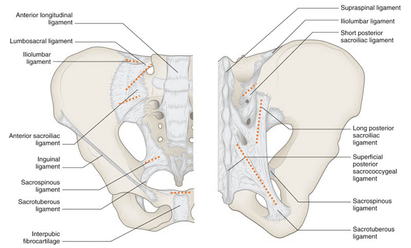
Ligaments of Pelvis
Inherent stability of the pelvis is provided by ligaments. The 3 groups of ligaments are: 1. Sacrum to Pelvis: Sacroiliac ligamentous complex: is divided into posterior (short and long) and anterior ligaments. Posterior ligaments provide most of the stability. Sacrotuberous ligament: runs from the posterolateral aspect of the sacrum and the…
PGMEE, MRCS, USMLE, MBBS, MD/MS
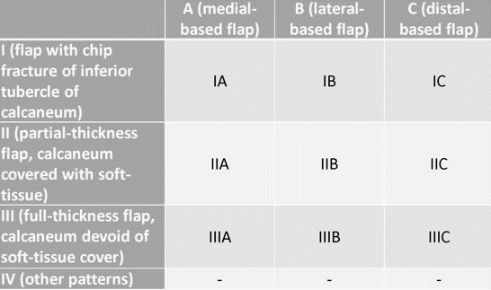
Heel Pad Avulsion Injuries
Special Anatomic Features of Heel Pad Heel pad form an almost fully contained cup-like structure consisting of skin overlying a shell of connective tissue within which fibrous septa ramify throughout the heel connecting the underlying periosteum of the calcaneus to the overlying reticular dermis, thereby anchoring skin to bone. Most…
PGMEE, MRCS, USMLE, MBBS, MD/MS
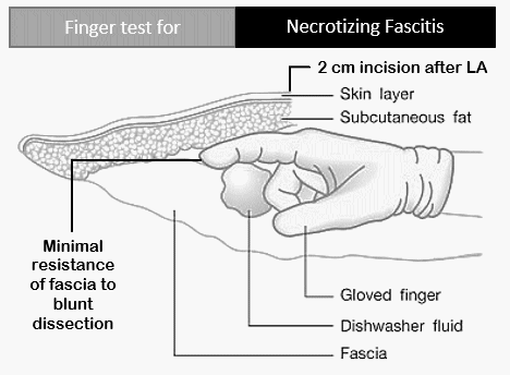
Differentiating Necrotizing Fasciitis from other soft tissue infections
Necrotizing fasciitis can be misdiagnosed in about 75% of the cases in the intial stage of the disease. The most consistent feature of early necrotizing fasciitis is the pain out of proportion to swelling or erythema. Other features helping to differentiate from other soft tissue infections are: Tenderness extending beyond…
PGMEE, MRCS, USMLE, MBBS, MD/MS
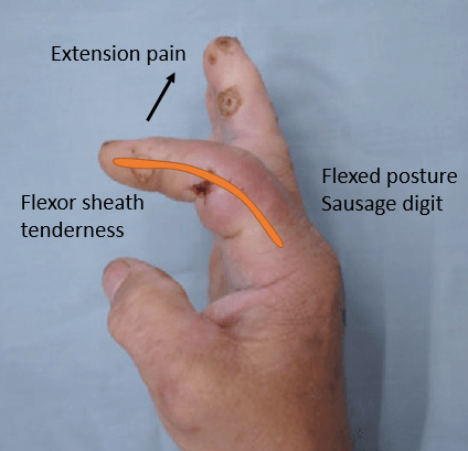
Kanavel Sign for Pyogenic Flexor Tenosynovitis
1. Exquisite tenderness over the course of the sheath, limited to the sheath Present in 64% cases Late sign of proximal extension of pyogenic tenosynovitis Most important sign as described by Kanavel 2. Flexion of the finger (‘hook’ sign) Present in 69% cases 3. Exquisite pain on extending the finger,…
PGMEE, MRCS, USMLE, MBBS, MD/MS
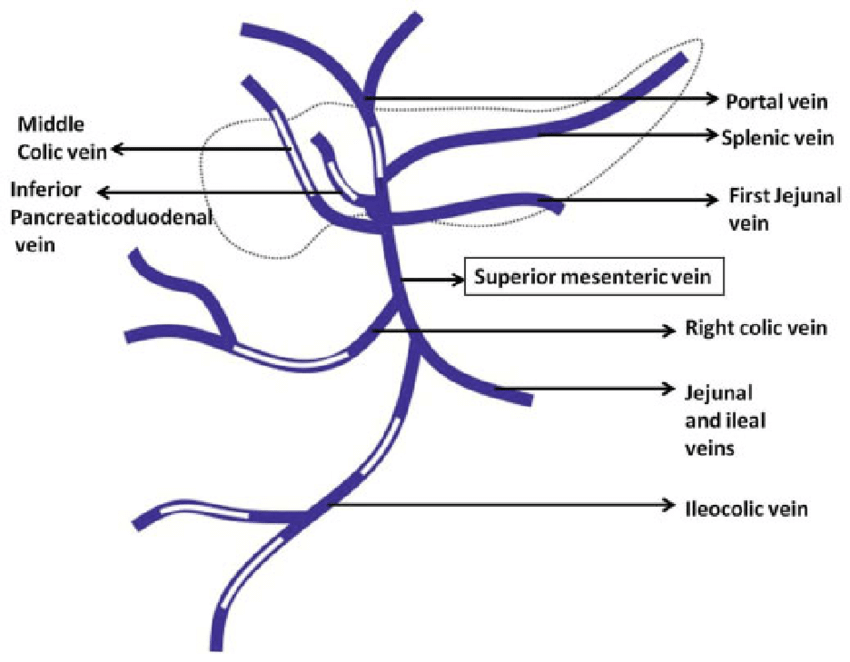
Portal Vein : Tributaries and Portocaval Anastomoses
Origin: Hepatic Portal Vein is formed by the union of Splenic vein and Superior mesenteric Vein behind the neck of pancreas at L1 vertebral level. Termination: The portal vein terminates by branching into right branch (entering right lobe of liver) and left branch entering (left lobe of liver). Parts: Tributaries: Points to…