Skip to content
Epomedicine

Mnemonics, Simplified Concepts & Thoughts

Epomedicine

Mnemonics, Simplified Concepts & Thoughts

Surgical Anatomy of Diaphragm

Dr. Manoj Bhandari, MS General Surgery Resident, Jan 20, 2024Jan 20, 2024

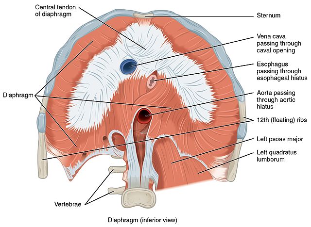
Diaphragm is a thoracoabdominal organ which is musculotendinous. It is the primary muscle of respiration. 

Extension:

  • Sternal – xiphoid process and transverse abdominis aponeurosis
  • Costal – lower 6 ribs and costal cartilage
diaphragm inferior view
OpenStax, CC BY 4.0 https://creativecommons.org/licenses/by/4.0, via Wikimedia Commons

There are 2 crura of diaphragm:

  • Right crus – L1 to L4
  • Left crus – L1 to L3

3 ligaments:

  • Lateral arcuate ligament 
  • Medial arcuate ligament 
  • Median arcuate ligament 

Quadratus lumborum muscle is related to lateral arcuate whereas psoas muscle is related to medial arcuate ligament.

Diaphragmatic openings
Patial T, Negi S, Thakur V. Hernia of Morgagni in the Elderly: A Case Report. Cureus. 2017 Aug 8;9(8):e1549. doi: 10.7759/cureus.1549. PMID: 29018646; PMCID: PMC5630465. Available via CC-BY-3.0.

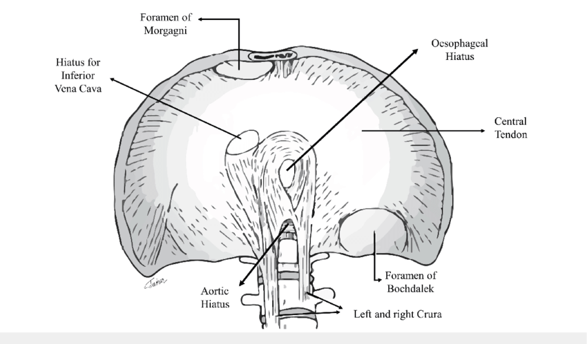
The foramen of Morgagni shown in the above picture (anteromedially) should obliterate normally. If it does not do so it results in congenital diaphragmatic hernia.

3 openings:

  • IVC at T8 level
  • Esophageal hiatus at T10 level
  • Aortic opening at T12 level

Other structures passing through different openings in diaphragm:

  • Caval hiatus: IVC and branches of right phrenic nerve
  • Esophageal hiatus: esophagus, vagal trunks, esophageal branch of left gastric artery and left gastric vein
  • Aortic hiatus: aorta, thoracic duct, azygous and hemiazygos vein
  • Minor apertures (through crura): greater, lesser and least splanchnic nerves 
Thoracic Diaphragm : Mnemonics

Arterial supply:

diaphragm blood supply
Gharagozloo, F. and Meyer, M. (2022) Diaphragmatic Flap: Technique of Preparation and Indications for Use. World Journal of Cardiovascular Surgery, 12, 207-218. Available via CC-BY-4.0.
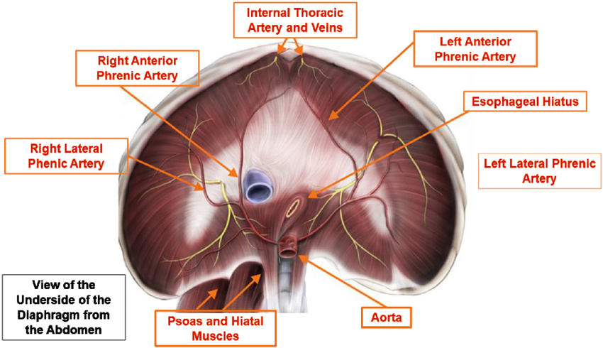
  • Major supply by right and left inferior phrenic artery (either as direct branch of aorta in 40% or from celiac trunk in 47%)
  • Superior phrenic artery supplies posterior part of diaphragm
  • Pericardiophrenic arteries- central area of diaphragm
  • Others: musculophrenic and lower intercostal arteries 

Venous drainage: 

  • Right inferior phrenic vein (IPV) directly drains into IVC
  • Left IPV either drains directly to IVC or can drain to left suprarenal vein which in turn drains into left renal vein then to IVC

Nerve supply:

  • Motor supply – right and left phrenic nerve which are divided as sternal, anterolateral and posterolateral branch 
  • Sensory supply:
    • Central portion: phrenic nerve 
    • Peripheral: lower 5 intercostal and subcostal nerve 

Embryological structures and their adult derivatives:

Embryonic structuresAdult derivatives
Septum transversumCentral tendon of diaphragm
Pleuroperitoneal membraneSmall peripheral part of diaphragm
Dorsal mesentery of esophagusCrura of diaphragm
Mesoderm of body wallLarge peripheral part of diaphragm external to parts derived from pleuroperitoneal membrane
Diaphragm and Body Cavities Development – Embryology Made Easy

Reference: Fischer’s mastery of surgery 8th edition

Dr. Manoj Bhandari
Dr. Manoj Bhandari, MS General Surgery Resident

He is an avid reader, guitar player, melodious singer and old songs lover. He has a passion for making medical knowledge accessible and comprehensive.

26 shares
  • Facebook23
  • Twitter
PGMEE, MRCS, USMLE, MBBS, MD/MS AnatomyGeneral SurgeryRespiratory system

Post navigation

Previous post
Next post

Related Posts

PGMEE, MRCS, USMLE, MBBS, MD/MS brachial plexus

Brachial Plexus Simplified with Mnemonics

Aug 19, 2016Dec 13, 2024

Components of Brachial Plexus Mnemonic: Randy Travis Drinks Cold Beer From proximal to distal, brachial plexus consists of: How are the roots formed? From the Ventral Rami of C5 to T1 spinal nerves. Extent and course: Intervertebral foramina to Transverse process to Interscalene triangle (bounded by anterior scalene and middle…

Read More
PGMEE, MRCS, USMLE, MBBS, MD/MS history and examination format

History and Physical Examination Format

Dec 5, 2017Dec 8, 2022

HISTORY 1. General Information: 2. Chief complaints: Example: Lower abdominal pain X 2 daysNausea and vomiting X 1 day 3. History of Presenting Illness: “OPQRST” for each symptoms Negative history: Treatment received for the complaint Review of systems: may or may not be related to chief complaint – include only…

Read More
PGMEE, MRCS, USMLE, MBBS, MD/MS

Paget’s Disease of bone : Mnemonic

Jul 20, 2022Jul 20, 2022

Synonyms: Osteitis deformans Collage of matrix madness Mnemonic: Sixteen “P”s 1. Paramyxovirus infection and inclusions in osteocalsts 2. Pain (most common symptom – localized fractures or nerve compressions) 3. Pelvis (most common site) 4. Polyostotic (more common; 85%) 5. Phases: Active (lytic, mixed, sclerotic) and Inactive 6. Plasma volume expansion…

Read More

Leave a Reply Cancel reply

Your email address will not be published. Required fields are marked *

This site uses Akismet to reduce spam. Learn how your comment data is processed.

Bhandari, M. Surgical Anatomy of Diaphragm [Internet]. Epomedicine; 2024 Jan 20 [cited 2026 Jun 5]. Available from: https://epomedicine.com/medical-students/surgical-anatomy-of-diaphragm/.

Pre-clinical (Basic Sciences)

Anatomy

Biochemistry

Community medicine (PSM)

Embryology

Microbiology

Pathology

Pharmacology

Physiology

Clinical Sciences

Anesthesia

Dermatology

Emergency medicine

Forensic

Internal medicine

Gynecology & Obstetrics

Oncology

Ophthalmology

Orthopedics

Otorhinolaryngology (ENT)

Pediatrics

Psychiatry

Radiology

Surgery

RSS Ask Epomedicine

  • What to study for Clinical examination in Orthopedics?
  • What is the mechanism of AVNRT?

Epomedicine weekly

  • About Epomedicine
  • Contact Us
  • Author Guidelines
  • Submit Article
  • Editorial Board
  • USMLE
  • MRCS
  • Thesis
©2026 Epomedicine | WordPress Theme by SuperbThemes