Skip to content
Epomedicine

Mnemonics, Simplified Concepts & Thoughts

Epomedicine

Mnemonics, Simplified Concepts & Thoughts

Obstacles to reduction in Developmental Dysplasia of Hip (DDH)

Dr. Sulabh Kumar Shrestha, MS Orthopedics, Nov 25, 2025Mar 23, 2026

Extra-articular obstacles: Secondary muscle shortening due to hip in subluxed/dislocated position

  1. Contracted adductor longus
  2. Contracted iliopsoas
    • Curves in front of the joint capsule
From: Farrow L et al., Surgical management for developmental dysplasia of the hip, Orthopaedics and Trauma, https://doi.org/
10.1016/j.mporth.2020.09.001
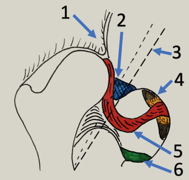
1. Anteromedial capsule; 2. Inverted limbus; 3. Iliopsoas tendon; 4. Pulvinar; 5. Ligamentum teres; 6. TAL

Intra-articular obstacles: Results in decreased volume of the acetabulum

  1. Thickened/elongated ligamentum teres
  2. Hypertrophied transverse acetabular ligament (TAL)
    • In crawling or walking child, caused by constant pull of ligamentum teres on its attachment at the base of the acetabulum
  3. Constricted anteromedial joint capsule
    • Most common and significant intra-articular obstacle
    • Caused by overlying contracted iliopsoas tendon
  4. Inverted and hypertrophied labrum (limbus)
    • Rare finding (seen only in teratologic dislocations and previously failed closed reductions)
  5. Pulvinar hypertrophy

Neo-limbus (acetabular roof hyaline cartilage): Neo-limbus is a hypertrophied ridge of fibrocartilage in the superolateral region of the acetabulum caused by pressure from the dislocated hip on this region and present in upto 98% cases of DDH in newborn. The palpable sensation of the femoral head gliding in and out of the acetabulum over this ridge of acetabular cartilage produces the Ortolani sign.

While some authors consider it can be an obstacle to reduction of hip, others have made following considerations:
a. Unlike limbus, it can invert or evert and is not an obstacle to reduction.
b. Neolimbus is not a fixed deformity and will spontaneously resolve once the femoral head has been reduced.
c. Neolimbus contains acetabular cartilage, and therefore epiphyseal cartilage, which is involved in the growth and development of the older child’s acetabulum and hence, should never be removed.

Further reading and references:

  1. Lovell and Winter’s Pediatric Orthopedics
  2. Landa J, Benke M, Feldman DS. The limbus and the neolimbus in developmental dysplasia of the hip. Clin Orthop Relat Res. 2008 Apr;466(4):776-81. doi: 10.1007/s11999-008-0158-y. Epub 2008 Mar 12. PMID: 18335297; PMCID: PMC2504652.
  3. Studer K, Williams N, Studer P, et al. Obstacles to reduction in infantile developmental dysplasia of the hip. Journal of Children’s Orthopaedics. 2017;11(5):358-366. doi:10.1302/1863-2548.11.170031

Mnemonic: PLACE

  1. Pulvinar
  2. Labrum (limbus), Ligaments (TAL, Ligamentum teres)
  3. Acetabular roof cartilage hypertrophy (Neolimbus) – controversial
  4. Capsule (anteromedial)
  5. Extra-articular (iliopsoas and adductor longus tendon)
dr. sulabh kumar shrestha
Dr. Sulabh Kumar Shrestha, MS Orthopedics

He is the section editor of Orthopedics in Epomedicine. He searches for and share simpler ways to make complicated medical topics simple. He also loves writing poetry, listening and playing music. He is currently pursuing Fellowship in Hip, Pelvi-acetabulum and Arthroplasty at B&B Hospital.

  • Facebook
  • Twitter
PGMEE, MRCS, USMLE, MBBS, MD/MS OrthopedicsPediatric hip

Post navigation

Previous post
Next post

Related Posts

PGMEE, MRCS, USMLE, MBBS, MD/MS

Surgical Site Infection (SSI) : CDC Definitions Simplified

Jan 18, 2021Jan 18, 2021

Superficial Incisional SSI Stab wound, pin site infection or stitch abscess alone is not considered an SSI. a. Time: Occurs within 30 days after any operative procedure (where day 1 = the procedure date) AND b. Site: Involves only skin and subcutaneous tissue of the incision AND c. Infection: At…

Read More
PGMEE, MRCS, USMLE, MBBS, MD/MS dural reflections and venous sinuses

Dural Reflections and Venous Sinuses

Aug 1, 2016Aug 22, 2023

Dura mater (pachymenix) is the outer meningeal layer consisting of: Dural Reflections These are the infoldings formed by the inner meningeal layer reflecting away from the fixed periosteral dural layer. Two vertical reflections – Separate the right and left hemisphere Two horizontal reflections Dural Venous Sinuses 1. Superior sagittal sinus:…

Read More
PGMEE, MRCS, USMLE, MBBS, MD/MS

Thyroid Gland Blood Supply : Mnemonics

Apr 29, 2024Apr 29, 2024

Arterial supply Arterial anastomosis Venous drainage The venous plexus lies beneath the true capsule (peripheral condensation of fibrous stroma of gland). Hence, during thyroidectomy, thyroid gland is removed along with the true capsule to avert the hemorrhage. False capsule (pre-tracheal fascia) is left intact. Thyroid Arteries and Laryngeal Nerves Mnemonic:…

Read More

Leave a Reply Cancel reply

Your email address will not be published. Required fields are marked *

This site uses Akismet to reduce spam. Learn how your comment data is processed.

Pre-clinical (Basic Sciences)

Anatomy

Biochemistry

Community medicine (PSM)

Embryology

Microbiology

Pathology

Pharmacology

Physiology

Clinical Sciences

Anesthesia

Dermatology

Emergency medicine

Forensic

Internal medicine

Gynecology & Obstetrics

Oncology

Ophthalmology

Orthopedics

Otorhinolaryngology (ENT)

Pediatrics

Psychiatry

Radiology

Surgery

RSS Ask Epomedicine

  • What to study for Clinical examination in Orthopedics?
  • What is the mechanism of AVNRT?

Epomedicine weekly

  • About Epomedicine
  • Contact Us
  • Author Guidelines
  • Submit Article
  • Editorial Board
  • USMLE
  • MRCS
  • Thesis
©2026 Epomedicine | WordPress Theme by SuperbThemes