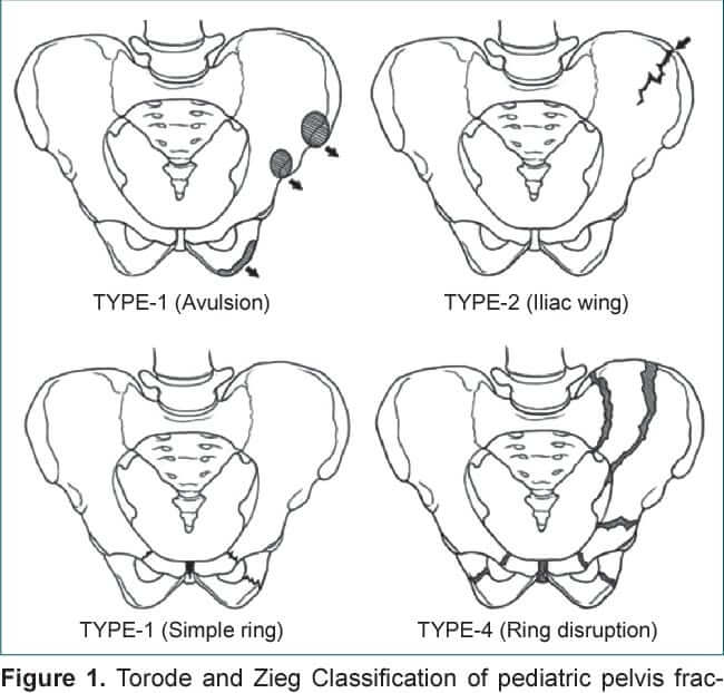
Before closure of triradiate cartilage (14 in boys and 12 in girls), pelvic bones are weaker than pelvic ligaments leading to more pubic rami and iliac wing fractures. After closure, they are more likely to sustain fractures of acetabulum, diastasis of pubic symphysis and SI joint separation. Hence, a pediatric…
Tag: Pediatric hip
Fellowship Blog
Revision Surgery After Failed Open Reduction in Developmental Dysplasia of the Hip
Re-dislocation rate after primary open reduction: 0-14% [1] Causes and timing of failure of primary surgery: Postoperative period Causes Immediate Approach related, Technical errors Delayed (After 4-6 weeks) Inadequate capsulorrhaphy, Inadequate immobilization Late Abnormal remodeling of acetabulum and femur Across multiple studies, inadequate soft tissue release – specifically the psoas…
Fellowship Blog
Approach to Neuromuscular Hip Dysplasia in Children with Cerebral Palsy : Principles of management and Long-term Radiographic Outcome
Dr. Sulabh Kumar Shrestha1, Dr. Rajendra Aryal2, Dr. Nitesh Raj Pandey2, Dr. Bibek Banskota21AKBEF Fellow, 2Unit-1 (Hip, Pelvi-acetabulum & Arthroplasty), B&B HospitalPoster presentation at Orthocon 2026 Introduction Progressive hip subluxation is a common orthopedic problem in children with cerebral palsy (CP), with strong correlation to Gross Motor Function Classification System…
PGMEE, MRCS, USMLE, MBBS, MD/MS
Obstacles to reduction in Developmental Dysplasia of Hip (DDH)
Extra-articular obstacles: Secondary muscle shortening due to hip in subluxed/dislocated position Intra-articular obstacles: Results in decreased volume of the acetabulum
Fellowship Blog
Classifications of Sequelae of Septic Arthritis of Hip in Children
Several radiological classifications have been developed to describe the long-term changes in the hip following septic arthritis and to help guide management. Hunka classification Choi classification Forlin and Milani classification Grade 1: Hips with the head or the femoral neck within the acetabulum Grade 2: Hips are dislocated Johari classification…
PGMEE, MRCS, USMLE, MBBS, MD/MS
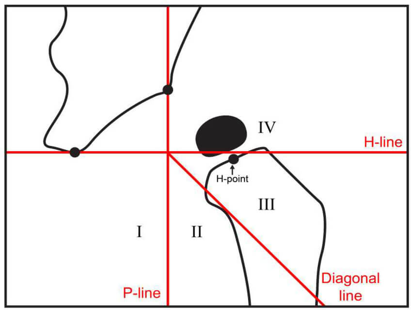
Tonnis and IHDI (International Hip Dysplasia Institute) Classification for DDH
IHDI method is a new radiographic classification of the severity of hip dislocation in DDH. It is based on the location of the midpoint of superior part of ossified metaphysis (H-point) relative to acetabulum. In contrast to Tonnis classification method, IHDI method: Tonnis classification: It is assessed according to the…
Fellowship Blog
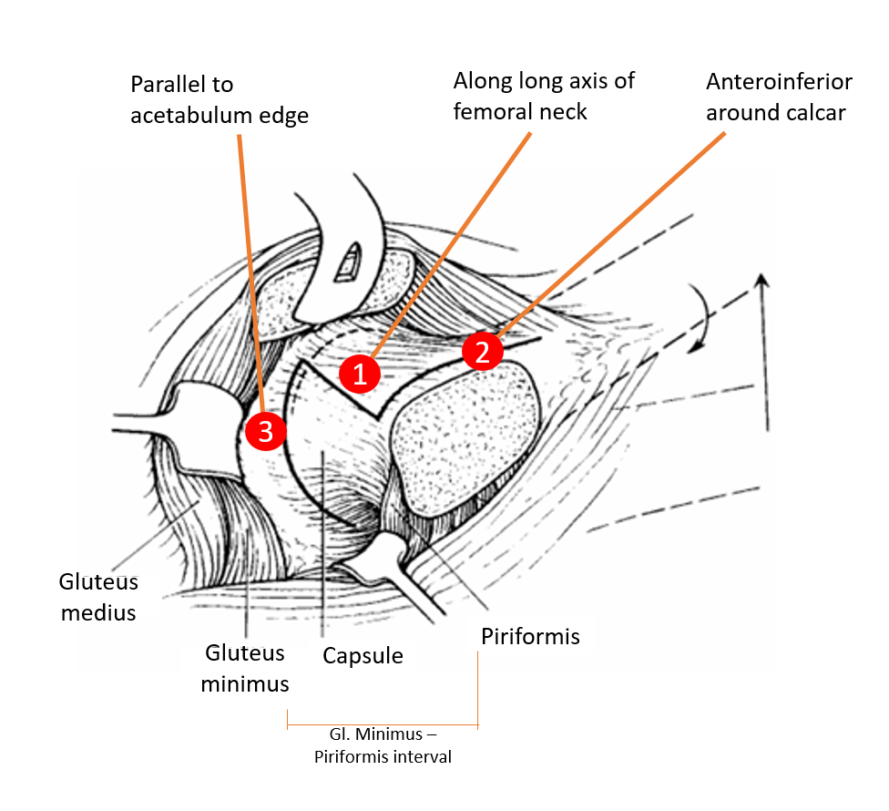
Safe Surgical Dislocation of Hip
Surgical Principles Relevant Course of MFCA Surgical Technique 1. Position: Lateral decubitus 2. Incision and approach: Traditional Kocher-Langebeck (KL) approach or Gibson interval (more anterior interval with posterior retraction of gluteus maximus muscle without violating it) 3. Trochanteric (Digastric slide) osteotomy: 4. Exposure: 5. Z-shaped Capsulotomy: 6. Hip Dislocation: 7….