While everyone is busy talking about the brachial plexus – lumbosacral plexus (the origin of nerves that supplies everything below the umbilicus) seems to be bit under-rated.
Formation of Lumbosacral Plexus
Ventral rami of L1-S4; has 2 components –
- Lumbar plexus (L1-L4) – forms within psoas major anterior to lumbar transverse process
- Sacral plexus (L4-S4) – forms anterior to piriformis muscle
Only in the lumb0sacral plexus, these anterior rami further divide into 2 divisions – 1 anterior and 1 posterior except:
- L4: Splits into 4 divisions – 2 anterior and 2 posterior
- S3: Doesn’t divide
Lumbar Plexus
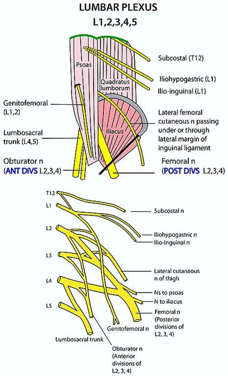
Formed from ventral rami of L1-L4.
Course: The nerves of the lumbar plexus exit the spine just anterior to the quadratus lumborum muscle and travel without and within the psoas muscle.
2 nerves with single root value: L1
1. Iliohypogastric nerve: Pierces the internal oblique muscle and runs between the internal and external oblique.
- Lateral cutaneous branches: pierces internal and external oblique 5 cm behind ASIS anterior superior iliac spine and just above the iliac crest – supplies skin on the anterior part of gluteal region.
- Anterior cutaneous branches: pierces aponeurosis of external oblique, 2 cm medial to ASIS – supplies skin over pubic bone (mons pubis).
2. Ilioinguinal nerve: Runs just inferior to iliohypogastric nerve, between transverse abdominis and internal oblique muscles.
- Pierces internal oblique 2 cm just below and medial to ASIS, runs below external oblique and inguinal canal with the spermatic cord and ends 2 cm lateral to pubic tubercle.
- Supplies mons pubis and and anterior aspect of labia majora; skin of anteromedial thigh and surrounding musculature.
2 nerves with double root values
L1, L2: Genitofemoral nerve
- Genital branch –
- Sensory: Skin of scrotum and labia majora
- Motor: Cremaster muscle
- Femoral branch – Skin on the antero-middle aspect of upper thigh
L2,L3: Lateral femoral cutaneous nerve
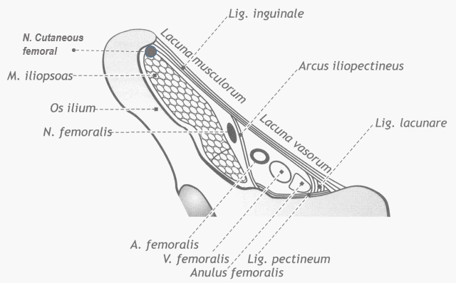
- Exits through lacuna musculorum (lateral compartment of thigh just below the inguinal ligament) along with iliopsoas muscle and femoral nerve.
- Supplies skin of anterolateral thigh.
2 nerves with triple root values: L2, L3, L4
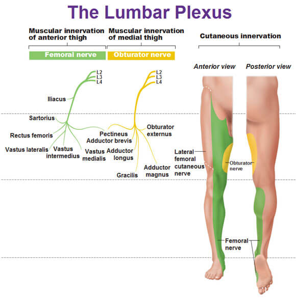
1. Anterior division: Obturator nerve
- Emerges from the medial border of psoas major and enters obturator canal with obturator artery and vein.
- Anterior branch: descends between adductor longus and adductor brevis
- Posterior branch: descends between adductor brevis and adductor magnus.
- Sensory: Medial aspect of thigh
- Motor: Hip adductors, Obturator externus
2. Posterior division: Femoral nerve
- Courses through psoas muscle, then iliopsoas groove and underneath the midpoint of inguinal ligament into the lacuna musculorum just medial to iliopsoas and lateral to femoral vessels.
- Divides into muscular and cutaneous branches in femoral triangle.
- Sensory: Anterior part of thigh and anteromedial part of leg.
- Motor: Hip flexors (Iliopsoas, Pectineus, Sartorius and Quadriceps femoris)
- Runs down as saphenous nerve.
A mnemonic is very popular: I Got Lunch (Laid) On Friday. Remember the progression of the lumbar root values:
- 1 – Ilioinguinal and Iliohypogastric nerves
- 1,2 – Genitofemoral nerve
- 2,3 – Lateral femoral cutaneous nerve
- 2,3,4 – Obturator and Femoral nerve
Sacral Plexus
Formed from ventral rami of L4-S4; Contribution of L4-L5 is from Lumbosacral trunk.
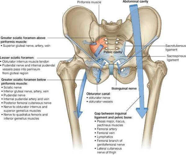
The nerves forming the sacral plexus converge towards the lower part of the greater sciatic foramen and unite to form a flattened band. The branches of the sacral plexus arise from anterior and posterior surfaces of this flattened band.
- Branches innervating the pelvis and perineum remain in the pelvis.
- Branches innervating the lower limb exit through greater sciatic foramen.
6 Branches Prior to Division of Sacral Roots
Mnemonic: All of these start with the letter “P”.
- S1,S2: Nerve to Piriformis
- S2, S3: Perforating cutaneous nerve – to medial part of buttock
- S1,S2,S3: Posterior femoral cutaenous nerve – To buttock and uppermost medial and posterior surfaces of thigh
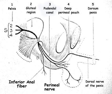
- S2,S3,S4:
- Pudendal nerve – leaves the pelvis through greater sciatic foramen but re-enters through lesser sciatic foramen and enters the pudendal or alcock’s canal (a fascial canal formed by splitting of obturator fascia on lateral wall of ischiorectal fossa).
- Inferior rectal nerve: Perianal skin and External anal sphincter
- Dorsal nerve of penis or clitoris: Accompanies dorsal artery of penis and clitoris and supplies skin of penis or clitoris and labia majora.
- Perineal nerve: Superficial and Deep perineal muscles and External urethral sphincter
- Parasympathetic pelvic splanchnic nerves (nervi erigentes) – Ascend to join inferior hypogastric plexus and together supply pelvic viscera.
- Pudendal nerve – leaves the pelvis through greater sciatic foramen but re-enters through lesser sciatic foramen and enters the pudendal or alcock’s canal (a fascial canal formed by splitting of obturator fascia on lateral wall of ischiorectal fossa).
- S4: Perineal branch of Pudendal nerve
Mnemonic: Branches of Pudendal nerve – PID (Remember – Pelvic Inflammatory Disease or PID can cause Pudendal neuralgia).
- Perineal nerve
- Inferior rectal nerve
- Dorsal nerve of penis or clitoris
In general, it supplies structures of perineum, is sensory to genitalia and gives muscular branches to perineal muscles, external urethral and anal sphincters (both are voluntary).
Another mnemonic: All these nerves arise from S1-S4 and have a vowel second latter after “P”. It progresses serially – except that the “e” and “i” are interchanged – so the sequence becomes a, i, e, o, u.
- Nerves with 2 roots:
- S1,S2 – Pirifromis nerve
- S2,S3 – Perforating cutaneous nerve
- Nerves with 3 roots:
- S1, S2, S3 – Posterior femoral cutaneous nerve
- S2, S3, S4 – Pudendal nerve and Parasympathetic pelvic splanchnic nerves
After Giving Anterior and Posterior Divisions
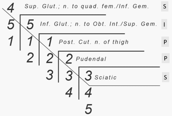
Here’s a great mnemonic video that assigns spinal levels to various nerves of sacral plexus.
| Anterior Division | Posterior Division | |
| L4-S1 (1st 3) | Nerve to Quadratus femoris and Inferior gemellus | Superior gluteal nerve
|
| L5-S2 (2nd 3) | Nerve to Obturator internus and Superior gemellus | Inferior gluteal nerve
|
| L4-S2 (All except S3 and S4) | Common peroneal (fibular) portion of Sciatic nerve
| |
| L4-S3 (All except S4) | Tibial portion of sciatic nerve
|
Sciatic nerve course:
a. Plexus: Largest branch of lumbosacral plexus (fromed from ventral rami of L4-S3)
b. Pelvis to Posterior gluteal region: Via greater sciatic foramen
c. Piriformis: Passes deep (anterior) to Piriformis (85% cases) and superficial to short external rotators
d. Posterior thigh:
- Runs superficial to adductor magnus
- Crossed obliquely by long head of biceps femoris
- Runs between semimembranosus and biceps femoris before entering popliteal fossa
- Supplies hamstring muscles
e. Popilteal fossa: At the apex of popliteal fossa, divides into:
- Peroneal nerve
- Wraps around the neck of fibula
- Divides into: Superficial peroneal nerve (supplies lateral compartment of leg) and Deep peroneal nerve or anterior tibial nerve (supplies anterior compartment of leg)
- Posterior tibial nerve
- Runs between superficial (triceps surae) and deep flexor compartment and supplies them
- Passes posterior to medial malleolus and divides into: medial (supply medial muscle group of foot except adductor hallucis and central muscle group) and lateral plantar nerves
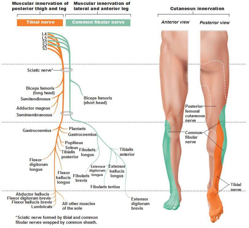
Sural nerve: It is formed by communication by sural communicating branches between the:
- Medial sural cutaneous branch (terminal branch of tibial nerve)
- Lateral sural cutaneous branch (terminal branch of common peroneal nerve)
From the mid calf down to the ankle the nerve courses subcutaneously along a line drawn from the mid-posterior popliteal fossa to just posterior to the lateral malleolus and thence under the malleolus and forward along the lateral aspect of the foot. It is purely sensory in function – supplies lateral foot and lateral lower ankle.
Lumbosacral Plexopathies or Lumbosacral Syndromes
Patterns of weakness usually help localize the “lesion” to a more specific area within the plexus. 1. Lumbar plexus lesions: weakness of hip flexion and adduction and/or knee extension.
- L2, L3, L4: Hip flexors and Knee extensors (Femoral nerve) and Hip adductors (Obturator nerve)
2. Lumbosacral trunk and upper sacral plexus lesions: foot drop (flail foot), and weakness of knee flexion or hip abduction.
- L4-S2: Common peroneal nerve (superficial peroneal – evertors of foot and deep peroneal – invertors and dorsiflexors of ankle and extensors of toes)
- L4-S3: Tibial nerve (knee flexors, dorsiflexors of toe, invertors of foot)
- L4-S1: Superior gluteal nerve (Pelvis stabilizer, i.e. hip extensors, abductors and internal rotators – lesion causes Trendelenburg sign)
- L5-S2: Inferior gluteal nerve (Hip extensor and Lateral rotator of thigh)
Patterns of sensory disturbance are less reliable given the difficult clinical delineation between dermatomal and named nerve sensory loss. In general, sensory disturbance involving:
- Anterior and medial thigh: lumbar plexus involvement
- Foreleg, dorsum of the foot, and posterior thigh: lumbosacral trunk and/or sacral plexus lesion
- Sensory loss on dorsum of foot except 1st webspace: Superficial peroneal nerve
- Sensory loss on dorsum of foot in 1st webspace and anterolateral leg: Deep peroneal nerve
- Sensory loss on sole: Tibial nerve

He is the section editor of Orthopedics in Epomedicine. He searches for and share simpler ways to make complicated medical topics simple. He also loves writing poetry, listening and playing music. He is currently pursuing Fellowship in Hip, Pelvi-acetabulum and Arthroplasty at B&B Hospital.

Wonderful!!!!.It takes lot of patience ,keen observation and time to make topics like these so easy.It’s been so helpful…..thank you so much