Skip to content
Epomedicine

Mnemonics, Simplified Concepts & Thoughts

Epomedicine

Mnemonics, Simplified Concepts & Thoughts

fetal circulation schematic

Fetal Circulation Made Easy

Epomedicine, Aug 8, 2016Jun 2, 2020

Following are the important features of fetal circulation:

1. Placenta plays the role of lungs; lungs are not functional:

  • Like pulmonary veins, left umbilical vein carries highly oxygenated blood from placenta to heart.
  • Like pulmonary artery, right and left umbilical arteries braing deoxygenated blood to placenta.

2. Mixing potentially occurs at 4 sites:

  • Left umbilical vein (Oxygen saturation ~80%) – Portal vein (Much bypassed by ductus venosus to Inferior Venacava)
  • Left umbilical vein – Inferior venacava – Portal vein (Oxygen saturation of IVC blood is ~65%)
  • Superior venacava – Right atrium (Much bypassed by Foramen ovale)
  • Pulmonary veins – Left atrium (Much bypassed by Foramen ovale and ductus arteriosus)
fetal circulation oxygenation

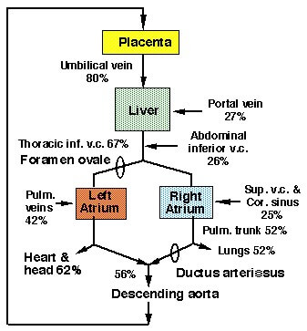
Oxygen saturation of blood in various vessels:

  1. Umbilical vein: 80%
  2. Abdominal IVC and portal vein: 28%
  3. Thoracic IVC: 67%
  4. Superior venacava: 25%
  5. Pulmonary veins: 42%
  6. Pulmonary trunk: 52%
  7. Umbilical arteries: 56%

Overview of Fetal Circulation:

fetal circulation schematic
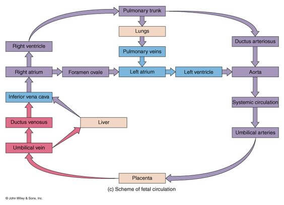
  1. Blood is oxygenated in the placenta.
  2. Highly oxygenated and nutrient-enriched blood returns to the fetus from the placenta via the left umbilical vein.
  3. Some blood enters liver sinusoids; most of the blood bypasses the sinusoids by passing through the ductus venosus and enters the inferior vena cava (IVC)
  4. From the IVC, blood enters the right atrium, where most of the blood bypasses the right ventricle through the foramen ovale to enter the left atrium.
  5. From the left atrium, blood enters the left ventricle and is delivered to fetal tissues via the aorta.
  6. Poorly oxygenated and nutrient-poor fetal blood is sent back to the placenta via right and left umbilical arteries.
  7. Some blood in the right atrium enters the right ventricle; blood in the right ventricle enters the pulmonary trunk, but most of the blood bypasses the lungs through the ductus arteriosus.

Fetal Combined Cardiac Output (CCO) Distribution:

Fetal cardiac output distribution

The 3 shunts of Fetal Circulation:

LocationShuntBetweenFunctionClosureRemnant
Pre-cardiac (Venous)Ductus VenosusLeft umbilical vein (Remember: Left is left and right regresses) and Inferior Venacava (IVC)Bypasses portal circulation


  • Left umbilical vein carries highly oxygenated blood (80%).
  • Prevents mixing with desaturated blood in portal circulation.

Has sphincter mechanism:

  • Closes and prevents overloading of heart during uterine contraction.
Functional: With umbilical cord clamping, venous return in the IVC decreases and reduction of flow through ductus venosus allows passive collapse.


  • occurs during the 1st minutes after birth and is not complete. An appreciable amount of the portal venous blood is shunted through this channel during the 1st hours and perhaps days of life.

Anatomical: Proliferation of the obliterating tissue in specially structured border strips by 15th to 20th days.

Ligamentum venosum
CardiacForamen OvaleLeft atrium and Right atriumBlood from IVC directly flows to Left atrium due to the valves in IVC i.e. Right atrium only acts as a conducting pathway


  • No mixing of deoxygenated blood from Superior Venacava (SVC)
  • Bypasses pulmonary circuit
Functional: With the 1st breath – septum primum is pressed agains septum secundum


  • Decreased right atrial pressure – interruption of placental blood flow
  • Increased left atrial pressure – Closure of ductus arteriosus (increased blood in pulmonary circulation)

Anatomical: Fusion of 2 apposed septa in about 1 year

Fossa ovale
Post-cardiac (Arterial)Ductus ArteriosusPulmonary artery and AortaBlood enters Right ventricle through the Right atrium from:


  • Superior Venacava
  • Inferior Venacava (some amount of blood is prevented from passing through foramen ovale by crista dividens, i.e. lower edge of septum secundum)

Pulmonary circuit is bypassed due to high resistance in pulmonary vessels – blood flows from pulmonary artery to aorta via ductus arteriosus.

Functional: Within 48 hrs if birth by muscular contraction


  • Increase in oxygen tension inhibits ductal smooth muscle voltage-dependent potassium channels, which results in an influx of calcium and ductal constriction.
  • PGE2 and PGI2 levels fall because of metabolism in the now functioning lungs and elimination of the placental source.


Anatomical: By intimal proliferation; takes 1-3 months.

Ligamentum arteriosus

Fate of Umbilical Vessels:

1. Umbilical arteries:

Functional closure: Immediately after birth due to thermal and mechanical stimuli and increased oxygen tension leading to constriction.

Anatomical closure: By fibrosis; takes 2-3 months

Remnants:

  • Distal part: Medial umbilical ligaments
  • Proximal part: Superior vesical arteries

2. Umbilical vein:

Obliterates shortly after birth like ductus venosus.

Remnant: Ligamentum teres hepatis

13 shares
  • Facebook13
  • Twitter
PGMEE, MRCS, USMLE, MBBS, MD/MS AnatomyEmbryologyPediatricsPhysiology

Post navigation

Previous post
Next post

Related Posts

PGMEE, MRCS, USMLE, MBBS, MD/MS glycolysis and gluconeogenesis

Glycolysis and Gluconeogenesis: Mnemonics

Jan 27, 2016Oct 28, 2024

It is not necessary to memorize each and every step of the process. We will only look into the major events. A. Meaning: Glyco (Sugar) + Lysis (Breaking or splitting) B. Synonyms: Embden-Meyerhof Pathway (EM Pathway) C. Site: Cytoplasm D. Enzyme basics: Kinase: Adds or removes phosphate from substrate (uses…

Read More
PGMEE, MRCS, USMLE, MBBS, MD/MS

Approach to a Child with Short Stature

Jan 15, 2022Jan 15, 2022

Definition of Short Stature Short stature refers to the height below 3rd Centile or 2 Standard Deviations (-2 SD) or more below the mean height for chronological age and gender for the standard population. When height is >-3 SD it’s most likely pathological. Assessment of Short stature 1. Accurate height…

Read More
PGMEE, MRCS, USMLE, MBBS, MD/MS

Thoracic Diaphragm : Mnemonics

Aug 7, 2023Aug 7, 2023

Embryology Mnemonic: Several Parts Build Diaphragm Embryonic components Parts of Diaphragm 1. Septum transversum Tendinous portion (Central tendon) 2. Pleuroperitoneal folds Muscular portion 3. Body wall muscles Muscular portion 4. Dorsal mesentery of esophagus Crura 3 Major Openings of Diaphragm Mnemonics: 1. I Eat Apples 8, 10, 12 or 2….

Read More

Comments (3)

  1. Guri says:
    Sep 30, 2017 at 6:49 am

    Rally easy

    Reply
  2. Umair says:
    Jul 31, 2018 at 1:05 pm

    Thnx for making it easy and tidy

    Reply
  3. Dr says:
    Mar 5, 2024 at 11:29 am

    This is one of the most sorted flow chart …

    Reply

Leave a Reply to Guri Cancel reply

Your email address will not be published. Required fields are marked *

This site uses Akismet to reduce spam. Learn how your comment data is processed.

Pre-clinical (Basic Sciences)

Anatomy

Biochemistry

Community medicine (PSM)

Embryology

Microbiology

Pathology

Pharmacology

Physiology

Clinical Sciences

Anesthesia

Dermatology

Emergency medicine

Forensic

Internal medicine

Gynecology & Obstetrics

Oncology

Ophthalmology

Orthopedics

Otorhinolaryngology (ENT)

Pediatrics

Psychiatry

Radiology

Surgery

RSS Ask Epomedicine

  • What to study for Clinical examination in Orthopedics?
  • What is the mechanism of AVNRT?

Epomedicine weekly

  • About Epomedicine
  • Contact Us
  • Author Guidelines
  • Submit Article
  • Editorial Board
  • USMLE
  • MRCS
  • Thesis
©2026 Epomedicine | WordPress Theme by SuperbThemes