Skip to content
Epomedicine

Mnemonics, Simplified Concepts & Thoughts

Epomedicine

Mnemonics, Simplified Concepts & Thoughts

Wound Strength

Epomedicine, Jul 5, 2025Jul 5, 2025

Wound strength is determined by both quantity and quality of collagen and an appropriate balance of deposition and breakdown. The amount of collagen in a wound plateaus at several weeks post-injury.

Timeline:

1. First few weeks: Strength increases rapidly, reaching about 20% of pre-injury strength at 3 weeks. 

2. Around 6 weeks: Wound strength increases significantly, nearing about 80% of the original strength. 

3. 3 months: Wound strength reaches approximately 80% of its original strength. 

4. Beyond 3 months: Strength continues to increase slowly, but the wound area will likely never be as strong as the original skin, typically reaching about 70-80%. 

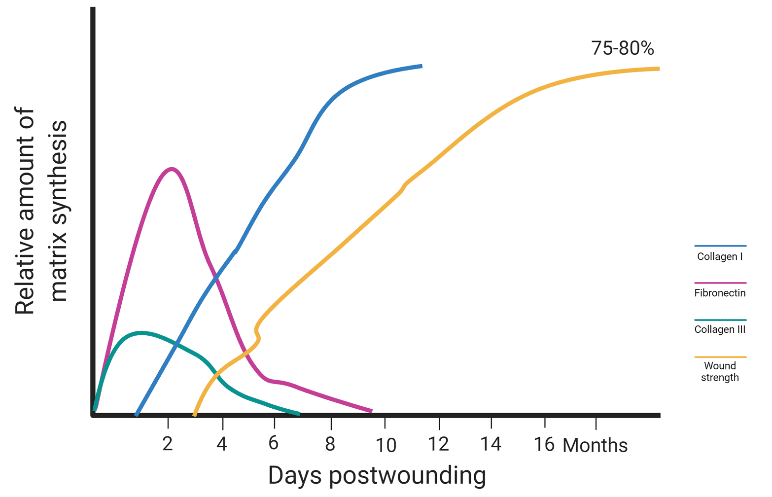
The tensile strength of the wound gradually increases over time. At 3 weeks, the wound has achieved 20% of its full strength. A wound’s maximum tensile strength peaks at 3 months, where it reaches at 80% of its pre-injury level.

  • Facebook
  • Twitter
PGMEE, MRCS, USMLE, MBBS, MD/MS General Surgery

Post navigation

Previous post
Next post

Related Posts

PGMEE, MRCS, USMLE, MBBS, MD/MS tympanogram interpretation

Understanding and Interpreting a Tympanogram

Aug 3, 2015

Relevant terms and definitions: 1. Tympanogram: plots compliance changes of the Tympanic Membrane (TM) versus air pressure in the EAC Y-axis shows pressure gradient and X-axis shows compliance change 2. Peak: point on the tympanogram that represents the point of maximum compliance, in which pressure of the external ear canal equals the pressure of the…

Read More
PGMEE, MRCS, USMLE, MBBS, MD/MS

Mediastinum : Simplified

Aug 7, 2023Sep 7, 2023

It is the region in thoracic cavity between the pulmonary cavities (covered by mediastinal pleura and excludes the lungs). Extent: Thoracic outlet (1st rib) to Diaphragm (T12) Superior mediastinum: Upper extent Thoracic outlet Contents (superficial to deep) 1. Glands (Thymus and Lymph nodes)2. 3 Veins (Brachiocephalic Lt. and Rt. and…

Read More
PGMEE, MRCS, USMLE, MBBS, MD/MS lung segments xray

Silhouette and Cervico-thoracic sign

Oct 30, 2016May 24, 2019

Synonyms: Obscured margin sign, Loss of outline sign Silhouette refers to the shadow and derived it’s origin from shadow papercuts done by Etienne de Silhouette. Principle of Silhouette sign On a normal Chest X-ray the well-defined borders of the heart and the domes of the diaphragm are visualised because the adjacent…

Read More

Leave a Reply Cancel reply

Your email address will not be published. Required fields are marked *

This site uses Akismet to reduce spam. Learn how your comment data is processed.

Pre-clinical (Basic Sciences)

Anatomy

Biochemistry

Community medicine (PSM)

Embryology

Microbiology

Pathology

Pharmacology

Physiology

Clinical Sciences

Anesthesia

Dermatology

Emergency medicine

Forensic

Internal medicine

Gynecology & Obstetrics

Oncology

Ophthalmology

Orthopedics

Otorhinolaryngology (ENT)

Pediatrics

Psychiatry

Radiology

Surgery

RSS Ask Epomedicine

  • What to study for Clinical examination in Orthopedics?
  • What is the mechanism of AVNRT?

Epomedicine weekly

  • About Epomedicine
  • Contact Us
  • Author Guidelines
  • Submit Article
  • Editorial Board
  • USMLE
  • MRCS
  • Thesis
©2026 Epomedicine | WordPress Theme by SuperbThemes