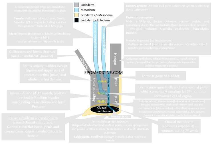
Cloaca literally meas a “sewer” – a coduit for carrying of drainage water and waste matter. The cloaca in human fetus have the same function, i.e. carries waste porducts in the from of urine and stool.
Cloaca: Common chamber for the hindgut and urinary systems.
- Formation of cloaca: Endodermal – incorporation of allantois into the hindgut during the 4th week of gestation due to folding of the embryo.
Cloacal membrane (plate): Membrane formed during 3rd week at the caudal end of the embryo from adhesion between epiblast and hypoblast cells. Later, it covers the cloaca.
- Rupture of cloacal membrane: During 7th week
Division of cloaca:
Mesodermal urorectal septum starts to divide cloaca from 4th week and reaches cloacal membrane to completely divide cloaca by 7 weeks:
- Anteriorly (connected to allantois): Urogenital sinus
- Posteriorly (connected to hindgut): Anorectal canal
- Apex of urorectal septum (in contact with cloacal membrane): Primitive perineum or perineal body
Fate of allantois:
- Early hematopoiesis (16th day i.e. 3rd week to 8 weeks)
- Complete obliteration by 15th week
Parts of Urogenital Sinus:
1. Cranially: Vesicle part
- Forms urinary bladder except trigone in both males and females.
- Trigone formation: By 8th week, ureteric bud invests into posterior part of urogenital sinus and forms trigone (epithelialized later)
- By 10th week: endodermal cells become single layer of cuboidal epithelium
- Subsequent weeks: additional cell layers appear, which begin to assume characteristics of differentiated urothelial cells
- During 12th week: muscularization of bladder – surrounding splanchnopleuric mesoderm differentiates to form the detrusor muscle lining the urothelium.
- Furhter maturation of bladder continues.
2. Middle: Pevlic part
- Forms prostatic and membranous urethra in males.
- The prostate develops from epithelial outgrowths form the prostatic segment of the urethra that grows into the surrounding mesenchyme.
- This outgrowth and branching start at week 10; by week 12, there are 5 groups of tubules that form the lobes of the prostate.
- Forms whole urethra in females.
- Vaginal plate:
- Paramesonephric ducts project into the dorsal wall of the pelvic part of cloaca and induce the formation of the sinovaginal bulbs.
- The sinovaginal bulbs fuse to form the solid vaginal plate, which canalizes and develops into the inferior two-thirds of
the vagina. - Completely canalized by 5th month.
3. Caudal: Phallic part (below the entrance of mesonephric ducts)
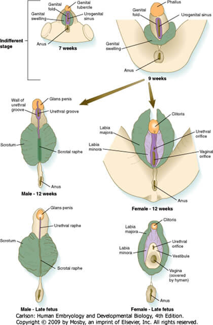
Indifferent stage: The primordia are indistinguishable up until about week 12.
- During 3rd week, mesenchyme migrate arounf the cloacal membrane through the primitive streak to form cloacal (urogenital) folds.
- Cranial to the cloacal membrane, the folds unite to form the genital tubercle.
- Genital (labioscrotal) swellings arise lateral to the urogenital folds.
- The overlying urogenital (cloacal) membrane becomes perforated in the urethral groove to form urogenital orifice so that as the urethral groove deepens, it is in communication with the phallic portion of urogenital sinus.
Differentiation of external genitalia:
| Genital tubercle | Urogenital folds | Genital swelling | |
| Male | Shaft and glans of penis
Corpora cavernosa |
Ventral shaft of penis
Corpora spongiosum Penile urethra (presence of androgen causes the genital folds to seal off and form a urethra) Urogenital sinus forms Bulbourethral glands of Cowper |
Scrotum
Scrotal raphe |
| Female | Clitoris | Open urogenital sinus (groove) forms Vestibule, Greater vestibular glands of Bartholin and Urethral and paraurethral glands of Skene.
Genital folds form labia minora |
Labia majora
Mons pubis |
During 4th month in males:
- Ectodermal ingrowth at tip of glans forms glandular epithelial plate which rapidly canalizes to form glandular urethra opening at the urethral meatus.
- The second invagination is circular and is called the preputial epithelial plate. Cleavage of this plate before birth separates the glans penis from the prepuce or foreskin.
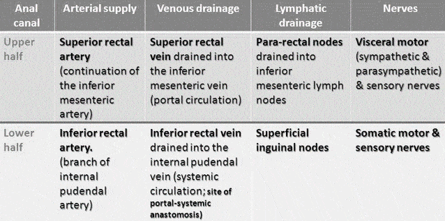
Hindgut: Part of the gut tube extending from the distal one-third of the transverse colon to the upper portion of the anal canal.
Proctodeum: Ectodermally lined pit that invaginates to form the lower third of the anal canal. Initially, this region is separated from the remainder of the anal canal by the anal membrane (once the posterior portion of the cloacal membrane), which breaks down to permit continuity between the two parts of the canal.
Pectinate line: Junction between hindgut and proctodeum – which also marks the site for anal membrane.
- In the adult, the pectinate line is located at the lower border of the anal columns.
Mesonephric and Paramesonephric ducts:
Above the cloaca –
1. Paramesonephric duct:
- Arises from genital ridge (intermediate mesoderm) lateral to Mesonephric duct:
- Female: Fallopian tubes, Uterus, Cervix, Superior 1/3 of vagina including fornices
- Vestigeal part: hydatid of Morgagni
- Male: Regress (influence of Mullerian Inhibiting Factor or MIF)
- Vestigenal remnant: Appendix testis
2. Mesonephric duct:
Urinary system: Ureteric bud gives collecting system (collecting ducts upto ureter)
Reproductive system:
- Male: epididymis, ductus deferens, seminal vesicle, and ejaculatory duct; efferent ductules (from mesonephric tubules)
- Vestigeal remnant of duct: Appendix epididymis
- Vestigeal remnant of tubules: Paradidymis (tubules)
- Female: regresses (no testosterone)
- Vestigeal remnant of duct: appendix vesiculosa, Gartner’s duct
- Vestigeal remnant of tubules: paraoophoron, epoophoron