Skip to content
Epomedicine

Mnemonics, Simplified Concepts & Thoughts

Epomedicine

Mnemonics, Simplified Concepts & Thoughts

choledochocele todani

Choledochal Cysts : Mnemonic for Todani Classification

Dr. Sulabh Kumar Shrestha, MS Orthopedics, Mar 2, 2018Mar 2, 2018


choledochocele todani

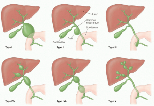
Todani Classification of Choledochal cysts

Mnemonic: Consider I is “Extrahepatic” and V is “Intrahepatic”, then –

  • Type I, II and III = Extrahepatic
  • Type IV = Extrahepatic + Intrahepatic
  • Type V = Intrahepatic

Most common: Type I

2nd most common: Type IV

Type I-III: Extrahepatic

Mnemonic: 123 EDC

Type I: Entire CBD dilated

Type II: Diverticulum

Type III: Choledochocele (Intraduodenal portion of CBD)

Type IV: Extrahepatic + Intrahepatic

Mnemonic: A for “And or Additional” and B for “Bunch”

Type IVa: Both intrahepatic AND extrahepatic cysts

Type IVb: Multiple extrahepatic cysts only

Type V: Intrahepatic cysts only (Caroli’s disease)

dr. sulabh kumar shrestha
Dr. Sulabh Kumar Shrestha, MS Orthopedics

He is the section editor of Orthopedics in Epomedicine. He searches for and share simpler ways to make complicated medical topics simple. He also loves writing poetry, listening and playing music. He is currently pursuing Fellowship in Hip, Pelvi-acetabulum and Arthroplasty at B&B Hospital.

56 shares
  • Facebook38
  • Twitter
PGMEE, MRCS, USMLE, MBBS, MD/MS Gastrointestinal systemGeneral SurgeryPediatrics

Post navigation

Previous post
Next post

Related Posts

PGMEE, MRCS, USMLE, MBBS, MD/MS hilum signs

Chest Xray – Approach to hilum

Jun 24, 2015Jun 24, 2015

Hilum in human anatomy refers to the depression where structures such as blood vessels and nerves enter an organ. The structures contributing to hilar shadows in a Chest X-ray are: Major: Pulmonary artery and veins Minor: Fat, Lymph nodes and Bronchial walls Normal Hilum: Position: Left hilum is slightly higher…

Read More
PGMEE, MRCS, USMLE, MBBS, MD/MS

Anatomy of Middle Ear with Clinical correlation

Jul 22, 2017Jun 12, 2020

The ear, along the evolution has modified structurally and functionally. In lower animals, they functioned as alarm systems to detect any sounds of the prey or predator so as to fix their vision and also maintain the balance of the body to prevent fall. In the course of evolution, this…

Read More
PGMEE, MRCS, USMLE, MBBS, MD/MS Mapleson breathing system

Mapleson Breathing Circuit Made Easy

Oct 17, 2016

Anatomy of Mapleson Breathing Circuit Basically, a mapleson breathing circuit consists of following parts: 1. Face mask (towards patient end) 2. Reservoir bag (towards operator end) Accommodates fresh gas flow during expiration acting as a reservoir available for the following inspiration. Acts as a monitor of patient’s ventilatory pattern during…

Read More

Comment

  1. vanscoyhair says:
    Mar 16, 2018 at 8:35 pm

    Thank you. Interesting.

    Reply

Leave a Reply Cancel reply

Your email address will not be published. Required fields are marked *

This site uses Akismet to reduce spam. Learn how your comment data is processed.

Pre-clinical (Basic Sciences)

Anatomy

Biochemistry

Community medicine (PSM)

Embryology

Microbiology

Pathology

Pharmacology

Physiology

Clinical Sciences

Anesthesia

Dermatology

Emergency medicine

Forensic

Internal medicine

Gynecology & Obstetrics

Oncology

Ophthalmology

Orthopedics

Otorhinolaryngology (ENT)

Pediatrics

Psychiatry

Radiology

Surgery

RSS Ask Epomedicine

  • What to study for Clinical examination in Orthopedics?
  • What is the mechanism of AVNRT?

Epomedicine weekly

  • About Epomedicine
  • Contact Us
  • Author Guidelines
  • Submit Article
  • Editorial Board
  • USMLE
  • MRCS
  • Thesis
©2026 Epomedicine | WordPress Theme by SuperbThemes