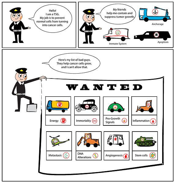
Today, we are going to talk only about the commonly mentioned tumor suppressor genes in the textbooks. This somewhat a “forced mnemonic” and may not be as effective and tedious to learn itself. If it works for you it’s well and good, if not find some other ways. General Rules…
Author: Dr. Sulabh Kumar Shrestha, MS Orthopedics
He is the section editor of Orthopedics in Epomedicine. He searches for and share simpler ways to make complicated medical topics simple. He also loves writing poetry, listening and playing music. He is currently pursuing Fellowship in Hip, Pelvi-acetabulum and Arthroplasty at B&B Hospital.
Blog
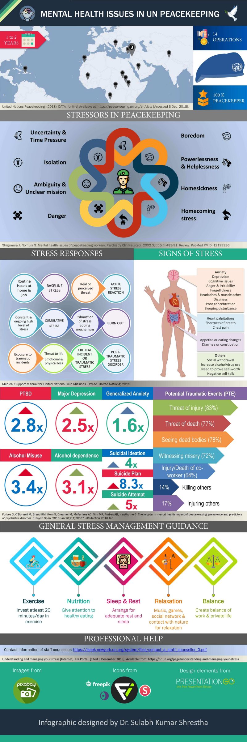
Mental Health Issues in UN Peacekeeping (Infographics)
This was done as a final presentation for the webinar course – “Social media and United Nations”. I have tried to create this infographic using microsoft powerpoint, free images and icons. Platform: Microsoft powerpoint Slide size: 30 inch X 10 inch Type of infographic: Statistical + Informational Title: Mental Health…
PGMEE, MRCS, USMLE, MBBS, MD/MS
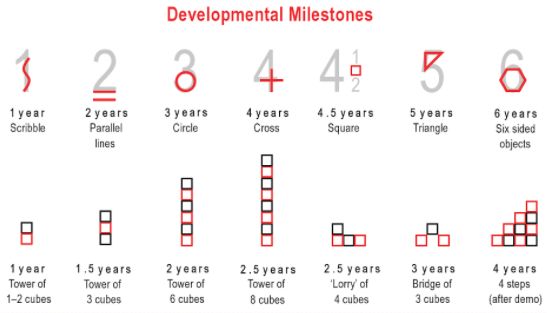
Developmental Milestones : Mnemonic
Principles of development Development proceeds from the head downward (cephalocaudal principle). Development proceeds from the center of the cody outward (proximodistal development). Development depends on maturation and learning. Development proceeds from the simple to the more complex. Growth and development is a sequential and continuous process. Growth and development proceed…
PGMEE, MRCS, USMLE, MBBS, MD/MS
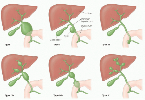
Choledochal Cysts : Mnemonic for Todani Classification
Todani Classification of Choledochal cysts Mnemonic: Consider I is “Extrahepatic” and V is “Intrahepatic”, then – Type I, II and III = Extrahepatic Type IV = Extrahepatic + Intrahepatic Type V = Intrahepatic Most common: Type I 2nd most common: Type IV Type I-III: Extrahepatic Mnemonic: 123 EDC Type…
Blog
Medical Journals of Nepal
History of scientific medical journal publication in Nepal In 2013, Journal of Nepal Medical Association (JNMA) celebrated golden jubilee. Founder Chief Editor of JNMA, Dr. Mrigendra Raj Pandey wrote, “My article was published in Indian Heart Journal way back in 1957 and became the first, which inspired me further and ultimately…
Emergency Medicine
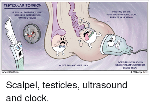
Testicular Torsion
Learning Objectives List the differential diagnosis for an acutely painful scrotum. Understand the anatomical basis of testicular torsion. List common presenting signs and symptoms of testicular torsion. Describe both initial and definitive management of testicular torsion. Explain why torsion is an emergent condition and discuss the time for salvage of…
Blog

Vancouver (NLM) Referencing Style : General rules of Citation
Vancouver style or NLM style of referencing is commonly used in medicine to cite other’s work in the text. General Rules 1. Reference numbers: Indicate the reference using arabic numerals inside [square] or (curved brackets) or as superscripts – use the same style throughout your scientific writing. Reference numbers must…
PGMEE, MRCS, USMLE, MBBS, MD/MS

History and Physical Examination Format
HISTORY 1. General Information: 2. Chief complaints: Example: Lower abdominal pain X 2 daysNausea and vomiting X 1 day 3. History of Presenting Illness: “OPQRST” for each symptoms Negative history: Treatment received for the complaint Review of systems: may or may not be related to chief complaint – include only…