
Strawberry in past has been mentioned in medicinal uses. This garden fruit is eponymoous to several important clinical signs in medicine. The list below is not a new one but a recompilation. Strawberry tongue: Surface of the tongue is coated with a thick white fur, through which protrude bright red…
Author: Epomedicine
PGMEE, MRCS, USMLE, MBBS, MD/MS
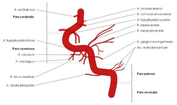
Internal Carotid Artery – Segments and Branches
7 segments of Internal Carotid Artery We have already discussed a mnemonic to remember the course of Internal Carotid Artery with the help of 2 horizontal “S” under the topic of Circle of Willis. C1 – Cervical segment C2 – Petrous (horizontal) segment C3 – Lacerum segment C4 – Cavernous…
PGMEE, MRCS, USMLE, MBBS, MD/MS
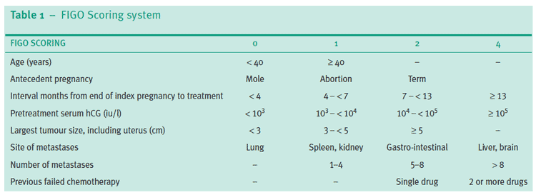
Gestational Trophoblastic Neoplasia (GTN) : Quick review
Follow-up protocol Post-Evacuation of Molar Pregnancy Weekly hCG measurements until hCG becomes undetectable Once hCG is undetectable, 2 further specimens should be obtained at weekly intervals Then monthly for 6 months and then Every 2 months for a further 6 months Diagnosis of GTN The diagnosis of GTN is made…
PGMEE, MRCS, USMLE, MBBS, MD/MS
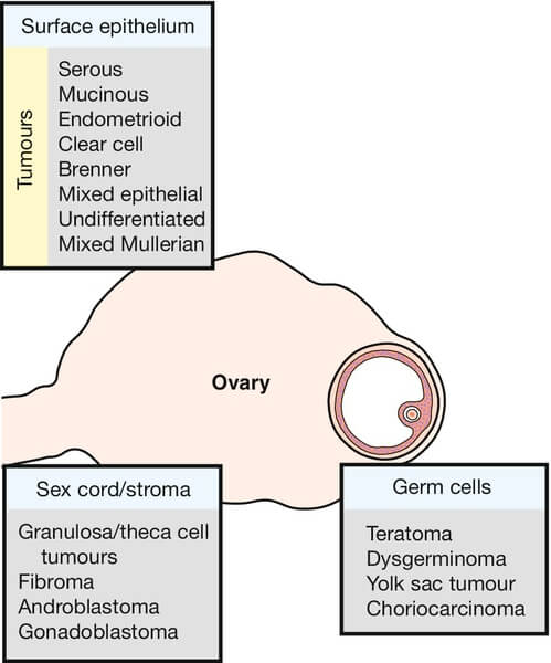
Pathology of Ovarian Tumors – Quick Review
The latest TNM and FIGO staging for Ovarian Cancer has been discussed earlier here. Here, we will discuss pathology of ovarian tumors in short and in a way thats easy to grasp. WHO Classification of Ovarian Tumors Cells of origin Surface coelomic epithelium Germ cells Sex cord, stromal cells Metastatic…
PGMEE, MRCS, USMLE, MBBS, MD/MS
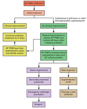
Ascitic Fluid Analysis : How to come to diagnosis?
We have already discussed about the practical essentials about Ascitic Paracentesis including absolute contraindications, site of needle entry and appropriate volume replacement. Laboratory analysis of ascitic fluid may provide answers to important clinical questions, as its composition varies depending on the underlying cause. Gross Special test WBC (/mm³) – most useful PMN…
PGMEE, MRCS, USMLE, MBBS, MD/MS
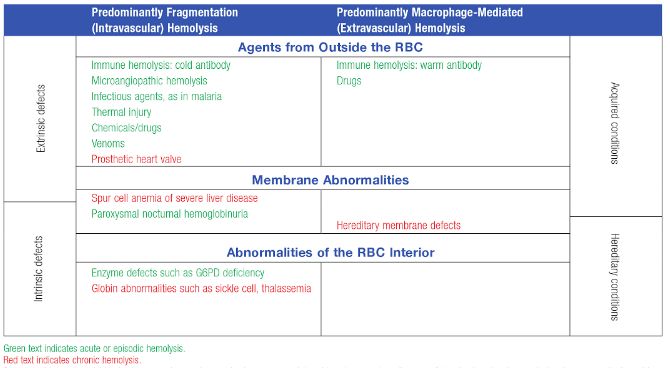
Hemolytic Anemia – Quick review
Although, we classify as intravascular and extravascular hemolysis, “diseases” don’t read the book. These disorders may be described as causing extravascular hemolysis, but your case may be the uncommon exception with intravascular hemolysis that was not mentioned. Diseases may cause anemia by both intravascular and extravascular hemolysis. Extravascular hemolysis typically…
PGMEE, MRCS, USMLE, MBBS, MD/MS
Understanding Red cell indices
Rule of 3s The measured hemoglobin concentration is 3 times the RBC count, and the calculated hematocrit is 3 times the Hb level. A significant deviation means artifacts in the value estimated or the RBCs are smaller or larger than the normal. HCt = 3 X Hb RBC count =…
PGMEE, MRCS, USMLE, MBBS, MD/MS
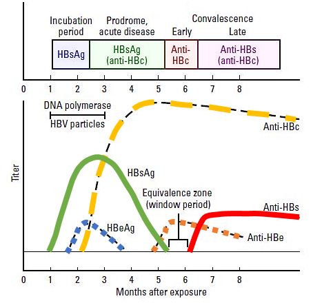
Interpreting Hepatitis B Serology in 5 Easy steps
Hepatitis B surface antigen (HBsAg) Appears during incubation period (1-6 months), 2-7 weeks prior to symptoms. Peaks when the patient is most ill. Becomes undetectable in 3-6 months. Indicates infection – recent or chronic. Hepatitis B surface antibody (anti-HBs or HBsAb) Arises once the acute disease has resolved. Sometimes, not…