Skip to content
Epomedicine

Mnemonics, Simplified Concepts & Thoughts

Epomedicine

Mnemonics, Simplified Concepts & Thoughts

anemia mcv-rdw

Understanding Red cell indices

Epomedicine, Feb 4, 2017

Rule of 3s

The measured hemoglobin concentration is 3 times the RBC count, and the calculated hematocrit is 3 times the Hb level. A significant deviation means artifacts in the value estimated or the RBCs are smaller or larger than the normal.

  • HCt = 3 X Hb
  • RBC count = Hb/3

The units

  1. Hematocrit: Percentage (%)
  2. MCV: fL (femtolitres i.e. 10^-15 litres)
  3. MCH: pg (picogram i.e. 10^-12 grams)
  4. MCHC: g/L

1 ml = 1000 cu.mm

1 L = 1000 ml

1 L = 1000000 cu.mm

Hb (g/dL) X 10 = Hb (g/L)
RBC indices

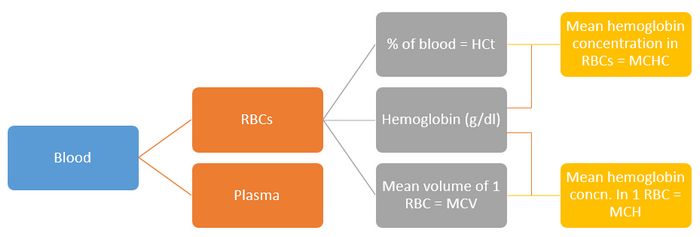
Hematocrit

In simple words, hematocrit is the percentage of your blood that is made up of Red Blood Cells (RBCs).

HCt = (Total blood volume – Total Plasma volume)/Total blood volume

Mean corposcular volume (MCV)

Mean volume of 1 RBC (Total RBC volume/Total number of erythrocytes or RBC)

MCV = (HCt X 10)/RBC count per Litre

Normal: 80-100 fL

Apply rules of 3 in the formula above: MCV = (3 X Hb X 10)/Hb/3 = 90. Hence, MCV is around 90 fL in average. Normal value is 90±10 fL.

HCt = MCV X RBC count X 0.1

Mean corposcular hemoglobin (MCH)

Mean hemoglobin concentration in 1 RBC (Hemoglobin concentration/Total number of erythrocytes or RBC)

MCH = (Hb X 10)/RBC count per Litre

Normal: 27-33 pg

Apply rules of 3 in the formula above: MCH = (3 X RBC count X 10)/RBC count = 30. Hence, MCH is around 30 pg in average. Normal value is 30±3 pg.

Mean corposcular hemoglobin concentration (MCHC)

Mean hemoglobin concentration in RBCs (Hemoglobin concentration/proportion of RBC)

MCHC = (Hb X 100)/HCt

Normal: 33-37 g/L

Apply rules of 3 in the formula above: (Hb X 100)/(3 X Hb) = 33. Hence, MCHC is around 33g/L in lower limit. Normal range is 35±2 g/L.

Interconversions

MCHC =  (MCH X 100)/MCV

MCH = (MCHC X MCV)/100

MCV = (MCH X 100)/MCHC

Red Cell Distribution Width (RDW)

Measure of anisocytosis (variability in RBC size) i.e. standard deviation of MCV

RDW = (Standard deviation of red cell volume X 100)/Mean cell volume

Normal range = 11 to 15%

Classification of Anemia based on MCV
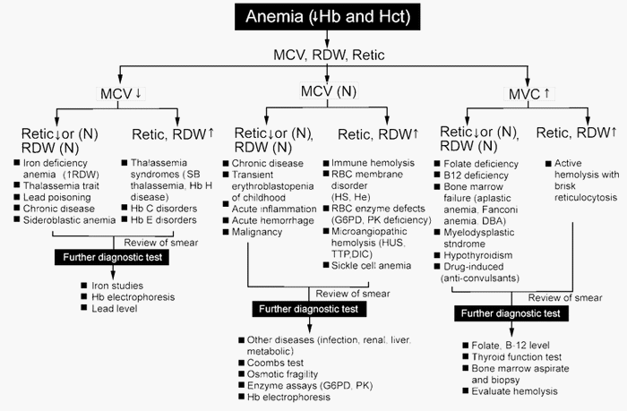
anemia mcv-rdw

MCV low (Microcytic)

Normal RDW

  • Thalasemmia trait
  • Anemia of chronic disease
  • HbH

High RDW

  • Iron deficiency
  • Beta-thalasemmia
  • Sickle/HbC trait

MCV normal (Normocytic)

Normal RDW

  • Normal
  • Anemia of chronic disease
  • Hemoglobinopathies
  • Hereditary spherocytosis
  • Transfusion
  • Chemotherapy
  • CLL, CML
  • Hemorrhage

High RDW

  • Mixed deficiency
  • Early iron or folate deficiency
  • Myelofibrosis
  • Sideroblastic anemia

MCV high (Macrocytic)

Normal RDW

  • Aplastic anemia
  • Pre-leukemia

High RDW

  • Folate deficiency
  • Vitamin B12 deficiency
  • Immune hemoglobin
  • Cold agglutinins
  • CLL
  • Facebook
  • Twitter
PGMEE, MRCS, USMLE, MBBS, MD/MS HematologyPathologyPhysiology

Post navigation

Previous post
Next post

Related Posts

PGMEE, MRCS, USMLE, MBBS, MD/MS

Kanavel Sign for Pyogenic Flexor Tenosynovitis

Sep 30, 2020Sep 30, 2020

1. Exquisite tenderness over the course of the sheath, limited to the sheath Present in 64% cases Late sign of proximal extension of pyogenic tenosynovitis Most important sign as described by Kanavel 2. Flexion of the finger (‘hook’ sign) Present in 69% cases 3. Exquisite pain on extending the finger,…

Read More
PGMEE, MRCS, USMLE, MBBS, MD/MS

Posterolateral corner (PLC) of knee

Oct 29, 2022Oct 29, 2022

Anatomy of PLC 3 major static stabilizers of PLC Lateral collateral ligament Popliteus tendon Popliteofibular ligament Other static stabilizers of PLC Lateral capsule thickening Arcuate ligament Fabellofibular ligament Dynamic stabilizers of PLC Biceps femoris Popliteus muscle Iliotibial band (ITB) Lateral head of gastrocnemius There are 2 “Y” shaped structures in…

Read More
PGMEE, MRCS, USMLE, MBBS, MD/MS internal capsule

Internal Capsule Simplified

Jul 30, 2016

Internal Capsule is a “boomerang” shaped (on horizontal section) and “funnel” shaped, i.e. tapering from superior to inferior (on sagittal section) white matter structure sandwiched between: Medially: Head of Caudate nucleus and Thalamus Laterally: Lenticular nucleus (Globus pallidus and Putamen) Parts of Internal Capsule 1. Anterior limb: Carries fibers to…

Read More

Leave a Reply Cancel reply

Your email address will not be published. Required fields are marked *

This site uses Akismet to reduce spam. Learn how your comment data is processed.

Epomedicine. Understanding Red cell indices [Internet]. Epomedicine; 2017 Feb 4 [cited 2026 Jul 28]. Available from: https://epomedicine.com/medical-students/understanding-red-cell-indices/.

Pre-clinical (Basic Sciences)

Anatomy

Biochemistry

Community medicine (PSM)

Embryology

Microbiology

Pathology

Pharmacology

Physiology

Clinical Sciences

Anesthesia

Dermatology

Emergency medicine

Forensic

Internal medicine

Gynecology & Obstetrics

Oncology

Ophthalmology

Orthopedics

Otorhinolaryngology (ENT)

Pediatrics

Psychiatry

Radiology

Surgery

RSS Ask Epomedicine

  • What to study for Clinical examination in Orthopedics?
  • What is the mechanism of AVNRT?

Epomedicine weekly

  • About Epomedicine
  • Contact Us
  • Author Guidelines
  • Submit Article
  • Editorial Board
  • USMLE
  • MRCS
  • Thesis
©2026 Epomedicine | WordPress Theme by SuperbThemes