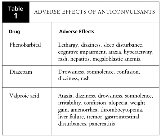
A) General consideration: Febrile seizures are seizures during fever occuring between 6 months and 5 years of age in absence of: CNS infections Abnormal neurologic findings Types of febrile seizure: Simple febrile seizure: Solitary Brief (< 15 minutes) Within 24 hours of onset of fever Generalized Tonic Clonic Seizure (GTCS)…
Tag: Pediatrics
Section Editor: Dr. Sujit Kumar Shrestha, MD Pediatrics, Fellowship Neonatology
Clinical Skills and Approaches

Nutritional History Made Easy
Energy Requirement 1. For a child with normal body weight: 100 Kcal/kg for 1st 10 kg Add 50 Kcal/kg for next 10 kg Add 20 Kcal/kg for body weight additional to 20 kgs 2. By age: For ≤ 1 year: 100 Kcal/kg/day Every additional years till puberty: Add 100 Kcal/year…
PGMEE, MRCS, USMLE, MBBS, MD/MS
Acute Diarrhea – Approach
There is no standard definition of diarrhea. Diarrhea may be defined with one or combination of the following criterion: Change in consistency of stool: Increased water-content Increase in freqency of stool: >3 times per day Increase in weight of stool: >200 grams per day or >10 grams/kg/day Among all these,…
PGMEE, MRCS, USMLE, MBBS, MD/MS
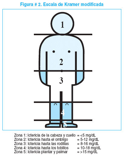
Neonatal Jaundice (NNJ) : Approach
Jaundice refers to accumulation of bilirubin in the epidermal tissues of the body, resulting in a yellowish tinge to the skin, sclera, and mucosa. Atleast 5 mg/dl of bilirubin level is required for clinically recognizing hyperbilirubinemia. A) Physiological Neonatal Jaundice: General consideration:
PGMEE, MRCS, USMLE, MBBS, MD/MS
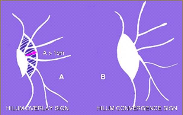
Chest Xray – Approach to hilum
Hilum in human anatomy refers to the depression where structures such as blood vessels and nerves enter an organ. The structures contributing to hilar shadows in a Chest X-ray are: Major: Pulmonary artery and veins Minor: Fat, Lymph nodes and Bronchial walls Normal Hilum: Position: Left hilum is slightly higher…
PGMEE, MRCS, USMLE, MBBS, MD/MS
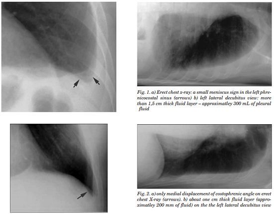
Chest X-ray – Pleural Effusion
Pleura is a mesothelial lined sac that envelopes the lungs and comprises of 2 membranous walls i.e. visceral pleura and parietal pleura that encloses pleural space filled with pleural fluid. Pleural space contains about 0.3 ml/kg body weight of pleural fluid. The pleura is not visible on a normal CXR…
Clinical Skills and Approaches

Skin signs of Dermatomyositis: Heliotrope rash, Grotton papules and Shawl sign
Dermatomyositis is a connective tissue disorder characterized by chronic inflammation of voluntary muscles and skin. It is more common in women and the age of onset is 50-70 years. A) Heliotrope Rash: It is a macular, confluent, purple or purple/red rash over both eyelids and periorbital tissue present with or…
PGMEE, MRCS, USMLE, MBBS, MD/MS
Chest X-ray: Alveolar vs Interstitial Disease
Interstitium is the scaffolding that supports the alveolar walls and surrounds both the alveoli and the terminal bronchioles. Neither alveoli nor interstitium is visible on a chest X-ray when normal. It is necessary to analyze whether the pattern of diffuse opacification in the lung field is alveolar or interstitial. Terms:…