Second week is the week of twos. In third week trilaminal germ disc is formed. And now, here I have attempted to fit the fourth week in development of embryo as the week of fours. Four folds of the embryo The embryo undergoes: Lateral folding Cranio-caudal folding It forms four…
Tag: Embryology
PGMEE, MRCS, USMLE, MBBS, MD/MS
Lung Development – Embryology Made Easy
Remember the mnemonic – “Every Premature Child Takes Air“. The development of lungs comprises of 5 distinct stages: Embryonic (3-8 weeks, i.e. embryonic period) Pseudoglandular (5-16 weeks) Canalicular (16-26 weeks) Terminal saccular (26-36 weeks) Alveolar (36 weeks to 40 weeks and continues to childhood) The first and last stages, i.e….
PGMEE, MRCS, USMLE, MBBS, MD/MS
Diaphragm and Body Cavities Development – Embryology Made Easy
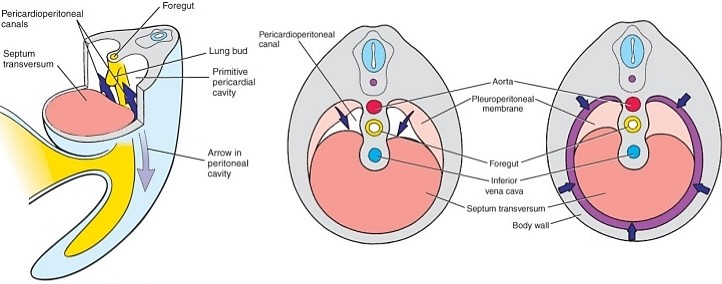
Let’s go back to the 4th week during the development of heart. The primitive intraembryonic coelom forms in the lateral and cardiogenic mesoderm during 4th week of development. Like the heart fields, the intraembryonic coelom has a horse-shoe configuration during this period: A thick mesodermal plate called “septum transversum” lies…
PGMEE, MRCS, USMLE, MBBS, MD/MS
Fetal Circulation Made Easy
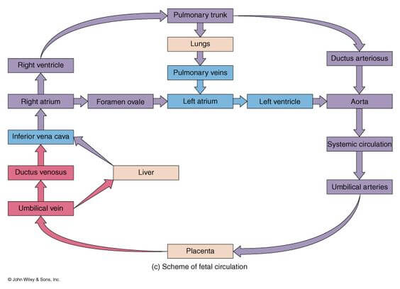
Following are the important features of fetal circulation: 1. Placenta plays the role of lungs; lungs are not functional: Like pulmonary veins, left umbilical vein carries highly oxygenated blood from placenta to heart. Like pulmonary artery, right and left umbilical arteries braing deoxygenated blood to placenta. 2. Mixing potentially occurs…
PGMEE, MRCS, USMLE, MBBS, MD/MS
Renal (Kidney) Development – Embryology Made Easy
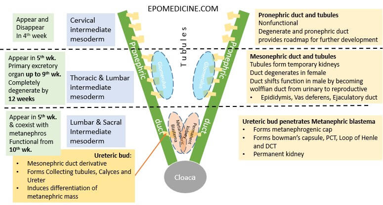
Kidney development occurs chronologically from cranial to caudal direction from urogenital ridge (intermediate mesoderm) in 3 different phases. Intermediate mesoderm → urogenital ridge (longitudinal elevation along dorsal body wall) → nephrogenic cord → Pronephros, Mesonephros and Metanephric mesoderm/blastema Remember the embryology of brain – from cranial to caudal, the primordial…
PGMEE, MRCS, USMLE, MBBS, MD/MS
Face and Palate Development – Embryology made Easy
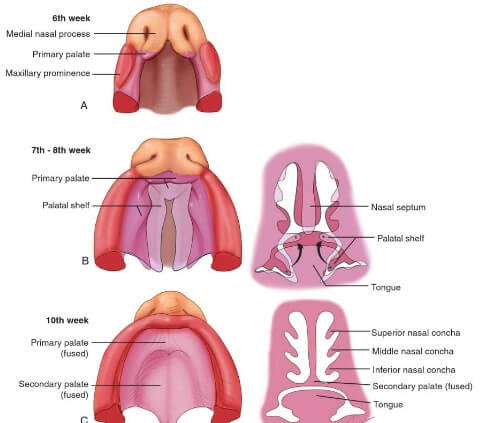
4 week embryo 5 mesenchymal prominences (facial primordia) appear in relation to the stomodeum (a depression in the surface ectoderm which marks the future mouth and oral cavity): Cranially: Frontonasal prominence (unpaired) Laterally: Maxillary prominence (paired; 1st pharyngeal arch) Caudally: Mandibular prominence (paired; 1st pharyngeal arch) 5 week embryo Localized…
PGMEE, MRCS, USMLE, MBBS, MD/MS
Heart Development – Embryology Made Easy
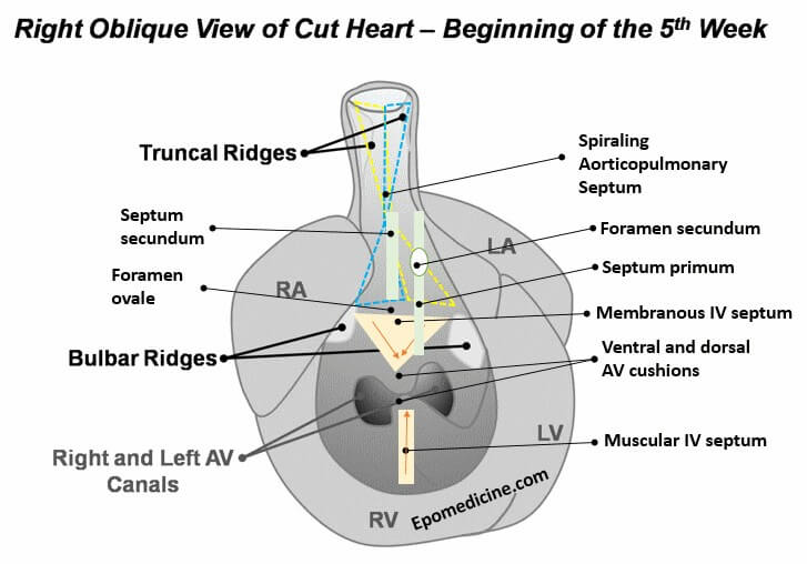
Heart Tube At the beginning of 4th week of development, heart is a continuous and valveless linear tube that resembles a chicken hung upside-down. It consists of 5 embryonic dilatation, that are destined to be the inflow and outflow tract and compartments of the hear without septum and valves. From cranial…
PGMEE, MRCS, USMLE, MBBS, MD/MS
Embryology Week 3: Trimalinar Germ Disc
In the Week 2, we had a bilaminar germ disc with 2 cavities (amniotic cavity and yolk sac) suspended in the chorionic (extraembryonic cavity) by a connecting stalk (future umbilical cord). In the 3rd week, the embryo will have 3 germ layers (ectoderm, mesoderm and endoderm) following a process called…