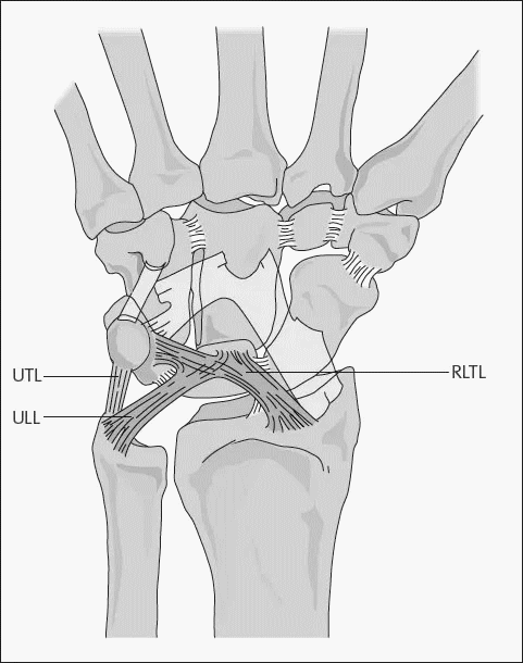
a. Interosseous: Extend deeply, directly between two bones Radioscapholunate (RSL) aka Ligament of Testut (neurovascular conduit to SL ligament) Scapholunate (SL) and Lunotriquetral (UL) – volar, dorsal and proximal fibrocartilaginous membrane components Capitohamate (CH) b. Palmar-proximal V: Converge as an “upside-down V” from the radius/ulna to lunate Radio-luno-triquetral (RLT) –…
Tag: Anatomy
PGMEE, MRCS, USMLE, MBBS, MD/MS
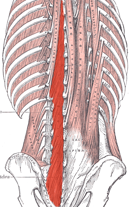
Muscles of Back – Simplified
A. Superficial Group (Appendicular group) Arise from vertebral column and attach to shoulder (assist in movement of limbs) a. Most superficial: Trapezius (From external occipital protuberance, ligamentum nuchae and spinous process C7-T12) Latissimus dorsi (From spinous process T7-T12, thoracolumbar fascia and iliac crest) b. Covered by trapezius: Levator scapulae (From…
PGMEE, MRCS, USMLE, MBBS, MD/MS
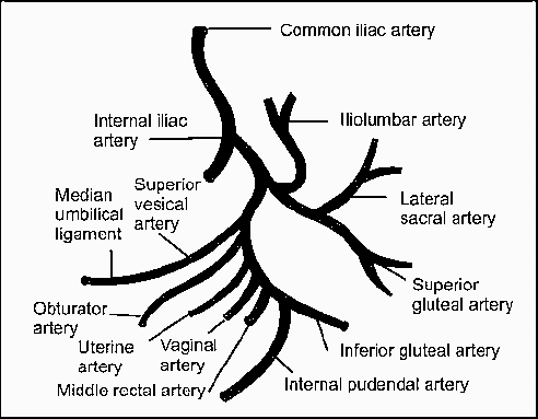
Internal Iliac Artery Anatomy : Simplified
Origin L5-S1 (common iliac artery bifurcation; anterior to SI joint) Course Extends down and posteriorly ~4 cm until superior margin of greater sciatic foramen and bifurcates into 2 trunks (in 60% cases) – Anterior trunk: continuation of the main artery towards ischial spine Posterior trunk: passes towards greater sciatic foramen…
PGMEE, MRCS, USMLE, MBBS, MD/MS
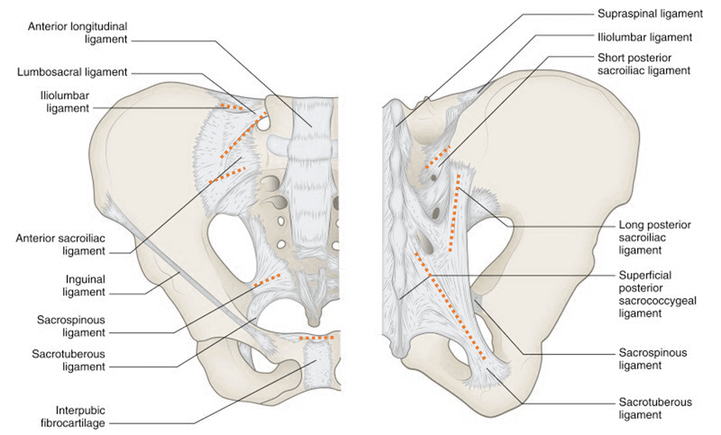
Ligaments of Pelvis
Inherent stability of the pelvis is provided by ligaments. The 3 groups of ligaments are: 1. Sacrum to Pelvis: Sacroiliac ligamentous complex: is divided into posterior (short and long) and anterior ligaments. Posterior ligaments provide most of the stability. Sacrotuberous ligament: runs from the posterolateral aspect of the sacrum and the…
PGMEE, MRCS, USMLE, MBBS, MD/MS
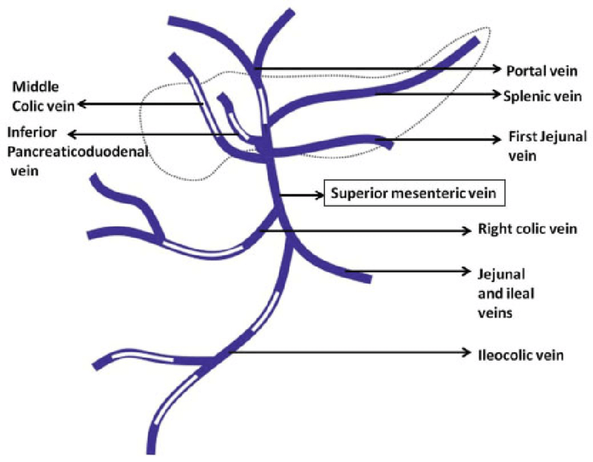
Portal Vein : Tributaries and Portocaval Anastomoses
Origin: Hepatic Portal Vein is formed by the union of Splenic vein and Superior mesenteric Vein behind the neck of pancreas at L1 vertebral level. Termination: The portal vein terminates by branching into right branch (entering right lobe of liver) and left branch entering (left lobe of liver). Parts: Tributaries: Points to…
PGMEE, MRCS, USMLE, MBBS, MD/MS
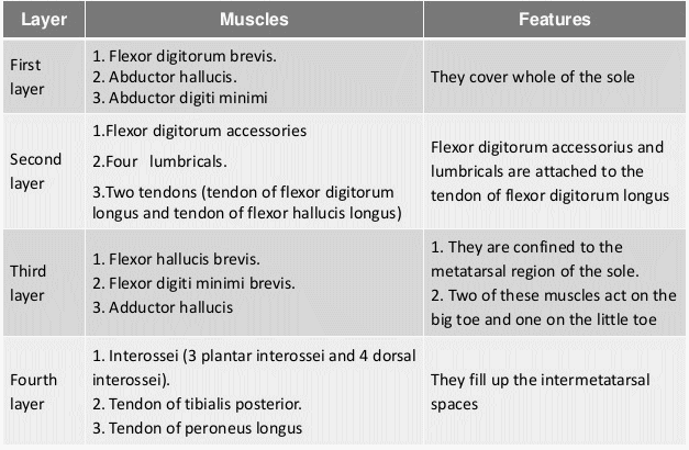
Foot muscles – Layers and Compartments
Deep to the plantar fascia, muscles of plantar foot exist in 4 different layers. Extensor digitorum brevis makes dorsal layer of foot and remaining 18 muscles and 4 tendons make the 4 layers of plantar aspect or sole of foot. In general: Layer 1 and 3: 3 muscles each that…
Clinical Skills and Approaches
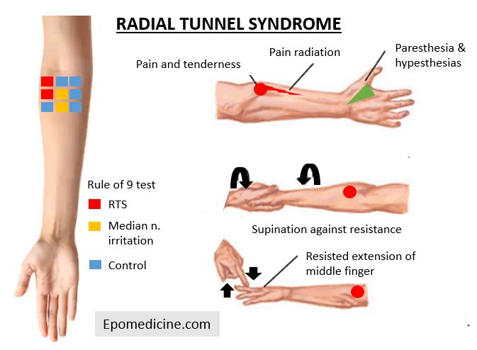
Radial Tunnel Syndrome (RTS) – Anatomy and Clinical Examination
Synonyms: Radial pronator syndrome, Treatment resistant lateral epicondylitis (tennis elbow) Anatomy of Radial Tunnel The anatomic radial tunnel (~5 cm) extends from the radial head to the inferior border of the supinator muscle. Mnemonic: FREAS Fibrous bands anterior to radiocapitellar joint Radial recurrent vessels (leash of Henry) ECRB medial edge…
PGMEE, MRCS, USMLE, MBBS, MD/MS
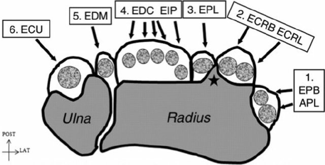
Wrist Extensor Compartments
The Extensor Zone VII (wrist) contains 6 extensor compartments comprising of 6 synovial sheath lined tunnels separated from each other by fibrous sheath. These compartments contain tendons of muscles that pass from forearm to hand. The number of tendons passinf thorugh the compartments (radial to ulnar) can be remembered using…