Skip to content
Epomedicine

Mnemonics, Simplified Concepts & Thoughts

Epomedicine

Mnemonics, Simplified Concepts & Thoughts

Radial Tunnel Syndrome (RTS) – Anatomy and Clinical Examination

Epomedicine, Jun 28, 2020Jun 28, 2020

Synonyms: Radial pronator syndrome, Treatment resistant lateral epicondylitis (tennis elbow)

Anatomy of Radial Tunnel

The anatomic radial tunnel (~5 cm) extends from the radial head to the inferior border of the supinator muscle.
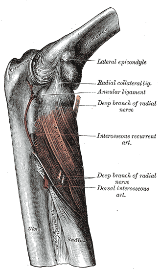
radial tunnel anatomy
Henry Vandyke Carter / Public domain

Mnemonic: FREAS

  1. Fibrous bands anterior to radiocapitellar joint
  2. Radial recurrent vessels (leash of Henry)
  3. ECRB medial edge
  4. Arcade of Frohse (proximal aponeurotic edge of supinator)
  5. Supinator (distal edge)

Anatomic areas 1-3 may engage sensory area while 4-5 doesn’t.

Anatomically, the radial nerve emerges from the supinator and then becomes the PIN (Posterior Interosseous nerve). Deep branch of radial nerve (DBRN) courses through the supinator to exit as the PIN at approximately 7.5 cm distal to the Radio-humeral joint.

Radial Nerve Anatomy : Course and Innervation

Diagnostic features

RTS may be confused with PIN syndrome but the main difference between the two is that RTS presents with sensory symptoms and any weakness is secondary to the pain while the PIN syndrome affects the motor portion of the nerve.

Radial tunnel syndrome tests

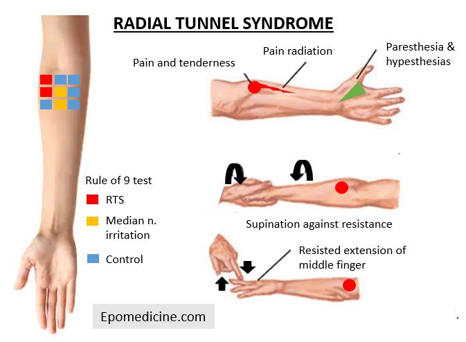
1. Patient may present with deep aching pain in dorsoradial proximal forearm which is worse at night and increases with lifting activities and forearm rotation and often accompanied by muscle weakness (due to pain rather than denervation).

2. Localized tenderness over the radial nerve 5 cm distal to lateral epicondyle.

3. Resisted long finger extension test – reproduces radial tunnel pain (some studies have considered as a pathognomic sign while in other studies there was no pain reproduction).

4. Resisted supination test – reproduces radial tunnel pain

5. Passive pronation with wrist flexion – passively stretches supinator muscle and increases pressure within radial tunnel to reproduce radial tunnel pain

6. Local anesthetic radial tunnel block – diagnostic if there is PIN palsy and pain is relieved.

7. Rule of Nine (RON) test – Subdivide the anterior, proximal forearm just distal to the elbow crease in to 9 regions arranged in a 3×3 grid. 3 medial regions without course of nerve are the control areas and expected to be free of pain and discomfort. Tenderness on the two proximal regions at the lateral column indicates radial nerve irritation. In the middle column, the two distal regions overlie the route of median nerve, and pain and tenderness in this area indicates a high level of median nerve irritation.

Reference: Moradi A, Ebrahimzadeh MH, Jupiter JB. Radial Tunnel Syndrome, Diagnostic and Treatment Dilemma. Arch Bone Jt Surg. 2015 Jul;3(3):156-62. PMID: 26213698; PMCID: PMC4507067.

44 shares
  • Facebook32
  • Twitter
Clinical Skills and Approaches AnatomyClinical examinationMusculoskeletal systemOrthopedics

Post navigation

Previous post
Next post

Related Posts

Clinical Skills and Approaches

Plantar fasciitis : Injection Technique

Aug 2, 2024Aug 2, 2024

Anatomy Plantar fascia arise from medial and lateral tubercles on the inferior surface of calcaneus. The lesion is invariably found at the medial head. Intersection technique 1. Position: Supine with knee flexed and hip externally rotated (figure of 4) 2. Landmark: Point of intersection of a line drawn parallel to…

Read More
Clinical Skills and Approaches

Communication Module for dealing with an Angry patient or relative

May 26, 2024May 26, 2024

The basic structure for communication skills remains the same. Recall the mnemonic P I CX3. Take into considerations of following points during consultation. Mnemonic: AARRR! Mnemonic Heading Comment A Acknowledge 1. Acknowledge the problem2. Acknowledge the concerns3. Acknowledge and validate the emotions of the patient/relative A Apologize and Empathize Apologize for the…

Read More
Clinical Skills and Approaches

Inverted Reflexes

Dec 8, 2022Dec 8, 2022

An inverted tendon reflex refers to the elicitation of the movement opposite to that normally seen when the reflex is elicited. Mechanism of Inverted Reflexes A lesion which simultaneously affects: This leads to 2 components: Mechanisms of hyperactive response: Types of Inverted Reflexes 1. Inverted supinator reflex: 2. Paradoxical triceps…

Read More

Leave a Reply Cancel reply

Your email address will not be published. Required fields are marked *

This site uses Akismet to reduce spam. Learn how your comment data is processed.

Pre-clinical (Basic Sciences)

Anatomy

Biochemistry

Community medicine (PSM)

Embryology

Microbiology

Pathology

Pharmacology

Physiology

Clinical Sciences

Anesthesia

Dermatology

Emergency medicine

Forensic

Internal medicine

Gynecology & Obstetrics

Oncology

Ophthalmology

Orthopedics

Otorhinolaryngology (ENT)

Pediatrics

Psychiatry

Radiology

Surgery

RSS Ask Epomedicine

  • What to study for Clinical examination in Orthopedics?
  • What is the mechanism of AVNRT?

Epomedicine weekly

  • About Epomedicine
  • Contact Us
  • Author Guidelines
  • Submit Article
  • Editorial Board
  • USMLE
  • MRCS
  • Thesis
©2026 Epomedicine | WordPress Theme by SuperbThemes