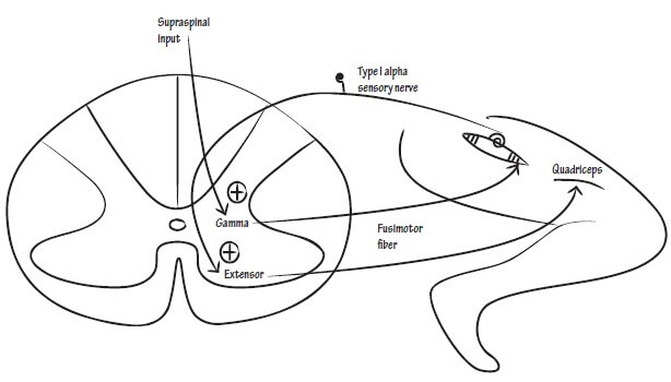
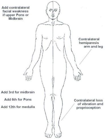
For the purpose of remembering the clinical manifestations of upper motor neuron lesion (UMNL) and lower motor neuron lesion (LMNL), a mnemonic has already been devised and discussed here. Now, it’s time to understand the anatomical and physiological basis of these manifestations. Upper Motor Neuron Lesion (UMNL) Syndrome Acute Manifestations…
Tag: Anatomy
PGMEE, MRCS, USMLE, MBBS, MD/MS
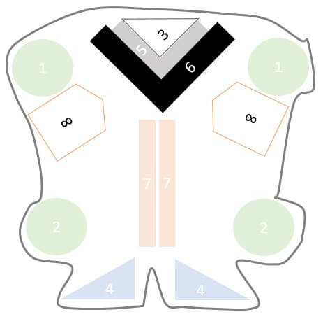
How to draw Medulla Oblongata Cross-section ?
Like in Midbrain and Pons: Corticospinal tract passes ventrally Ventricular system is located dorsally in midline Cranial nerve nuclei are located just anterior to the ventricle Medial longitudinal fasciculus is present around the center Another important thing to remember is that, the caudal medulla resembles “spinal cord“: Circular in shape…
PGMEE, MRCS, USMLE, MBBS, MD/MS

How to Draw Pons Cross-section ?
Pons in latin, refers to a “bridge”. Pons varolli is a part of brain-stem, that links thalamus with medulla oblongata. The cross-section of pons is similar to the midbrain as described earlier but few things must be kept in mind: The orientation of lemnisci in midbrain is more or less…
PGMEE, MRCS, USMLE, MBBS, MD/MS
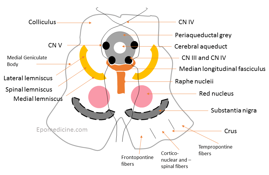
How to Draw Midbrain Cross-section ?
The cross-section of midbrain can be compared to the “upside down striped face of a red-eyed demon“. Using this analogy of a demon face, lets assign the structures found on the cross-section of midbrain: Ear = Crus cerebri Medial – frontopontine fibers Middle – corticonuclear and corticospinal tract Lateral –…
PGMEE, MRCS, USMLE, MBBS, MD/MS
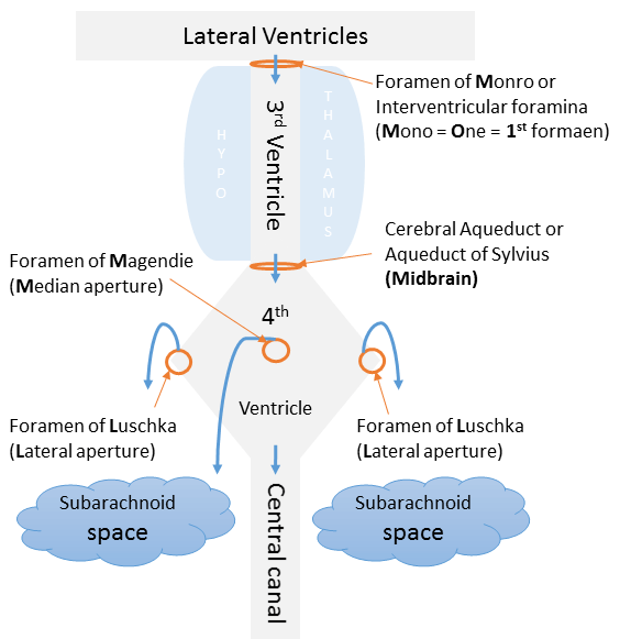
CSF Circulation Made Simple
Cerebrospinal Fluid (CSF) Production and Absorption CSF is produced by the choroid plexus that lines the ventricles. Choroid plexus = Infoldings of blood vessels of piamater + Modified ciliated ependymal cells Tight junctions of the choroid plexus cells form Blood-CSF barrier. CSF is reabsorbed by arachnoid granulations to enter dural…
PGMEE, MRCS, USMLE, MBBS, MD/MS
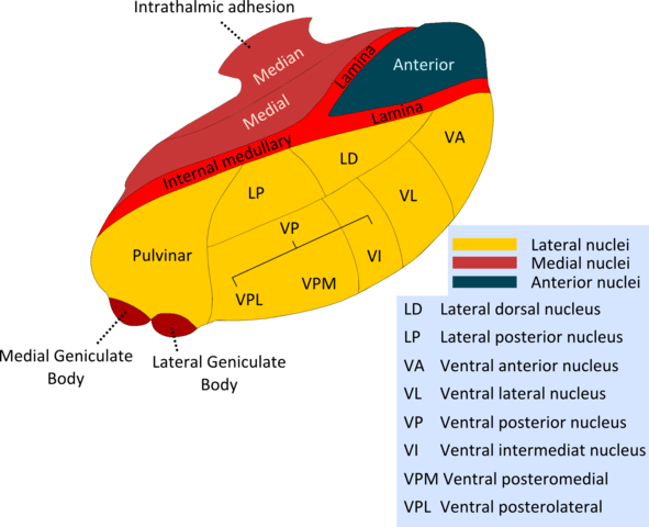
Thalamic Connections Mnemonic
Structure of Thalamus A vertical “Y” shaped white mater – internal medullary lamina divides thalamus into: In anatomical position: Pulvinar = Posterior end or posterior pole of thalamus Thalamic Connections Picture mnemonic Remember the schematic diagram drawn below showing important parts of thalamus in an anticlockwise fashion: Now, we assign…
PGMEE, MRCS, USMLE, MBBS, MD/MS
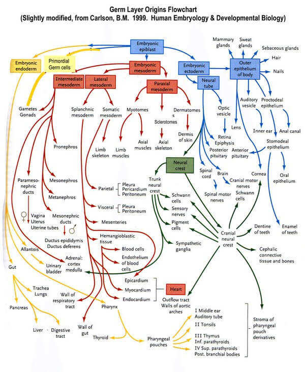
Germ Layer Derivatives – Mnemonics
Principle of Germ Layer Segmentation Ectoderm gives further rise to neuroectoderm and neural crest cells. Endoderm remains intact. Mesoderm gives further rise to paraxial mesoderm (somitomeres and 35 pairs of somites), intermediate mesoderm, and lateral mesoderm: General Rule for Germ Layer Derivatives Ectodermal derivatives: 1. Everything that makes you attractive: Skin, hair,…
PGMEE, MRCS, USMLE, MBBS, MD/MS
Medial Medullary (Dejerine’s) Syndrome : Anatomical basis mnemonic
As already discussed in the previous section about Lateral Medullary (Wallenberg) Syndrome: 6 “S” pass/lie on the Side (latetral) of Medulla Except the anteromedian part supplied by vertebral artery, rest of the medulla is supplied by PICA Let us now review the relevant anatomy and physiology of the medial portion…