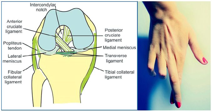
Mnemonic: Cross your long fingers over the index finger and superimpose this hand over your ipsilateral knee. This will help us to remember the orientation of the anterior cruciate ligament (ACL) and posterior cruciate ligament (PCL) of knee. Also remember the mnemonic “LAMP” which means Lateral ACL and Medial PCL….
Tag: Anatomy
PGMEE, MRCS, USMLE, MBBS, MD/MS
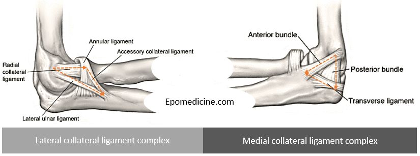
Elbow ligaments : Simplified Anatomy
Lateral collateral ligament complex Restraint to varus and posterolateral rotatory instability. Anatomy is more variable. LCL arises from lateral humeral condyle at a point through which the axis of rotation passes – it maintains a uniform tension throughout the arc of motion. Annular ligament is a “U” shaped ligament that…
PGMEE, MRCS, USMLE, MBBS, MD/MS
Shoulder Muscles Anatomy – Simplified
Proximal humerus Insertion of Rotato Cuffs Mnemonic: SIT-S a. Greater tubercle: Supraspinatus, Infraspinatus, Teres minor b. Lesser tubercle: Subscapularis Attachment of Other muscles Anteriorly and posteriorly the muscles attach on each side of the depressions (groove and sulcus). a. Anteriorly: Insertion of 3 muscles Mnemonic: Lady between 2 Majors Crest…
PGMEE, MRCS, USMLE, MBBS, MD/MS
Neuroanatomy Notes
This is a compilation of high yield topics on neuroanatomy with visual mnemonics and illustrations targeted for undergraduate medical students who find difficulty in conceptualizing the nervous system. Author: Dr. Sulabh Kumar Shrestha Key features: Covers clinically and academically important topics Schematic diagrams Visual mnemonics Useful for medical students, PG…
PGMEE, MRCS, USMLE, MBBS, MD/MS
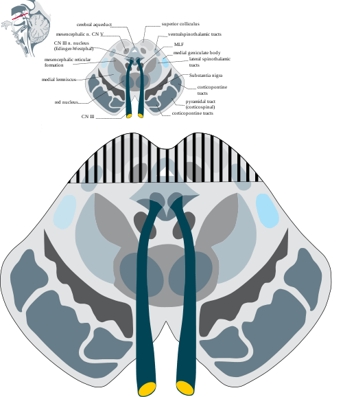
Parinaud Syndrome – Anatomical Basis
Synonyms: Dorsal midbrain sydnrome, Pretectal syndrome Main components of Parinaud syndrome: Vertical gaze palsy Convergence-retraction nystagmus Pupillary light-near dissociation Lid retraction (Collier’s sign) Anatomical basis of Parinaud syndrome: Site of lesion: Dorsal midbrain Close to cerebral aqueduct and pineal gland – hydrocephalus and pinealoma Arterial supply – posterior cerebral artery…
PGMEE, MRCS, USMLE, MBBS, MD/MS
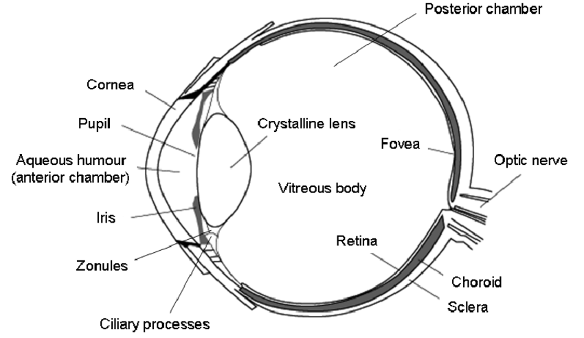
Embryology of Eye : Mnemonic
Surface ectoderm derivatives Mnemonic: LEVeL Lens Epithelium All ocular adnexa including mebomian gland and glands of Zeis and Moll Skin Cornea Conjunctiva Vitreous (portion) Lacrimal glands and drainage system Neuro-ectoderm (Optic vesicle and cup) derivatives Mnemonic: MORE Muscles of pupil (constrictor and dilator pupillae) Optic nerve Retinal pigment epithelium Epithelium…
PGMEE, MRCS, USMLE, MBBS, MD/MS
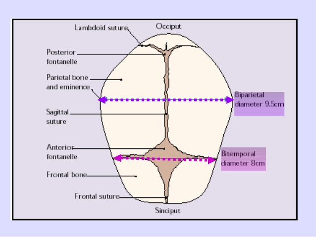
Fetal Skull Dimensions : Mnemonic
Anteroposterior Diameters Mnemonic: Larger the alphabets, larger the diameter (V>F>B) Diameters with “sub” prefix are smaller than counterpart without it. mento-Vertical: 14 cm SUB-mento-Vertical: 11.5 cm occipito-Frontal: 11.5 cm SUB-occipito-Frontal: 10.5 cm sub-occipito-Bregmatic: 9.5 cm sub-mento-Bregmatic: 9.5 cm Transverse Diameters Mnemonic: MTP (Medical Termination of Pregnancy) in increasing order. bi-Mastoid:…
PGMEE, MRCS, USMLE, MBBS, MD/MS
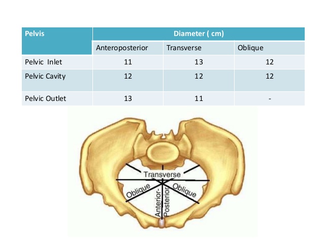
Pelvic Dimensions : Mnemonics
Shapes of the female pelvis Mnemonic: GAP (In order of most common to the least common) Only pelvis with AP diameter > Transverse diameter is Anthropoid pelvis. Mnemonic: ANthrOPoid Android and Anthropoid (AN) pelvis are common causes of occipito-posterior (OP) presentation. In platypelloid pelvis (broad and flat): Sub-public angle: It…