Skip to content
Epomedicine

Mnemonics, Simplified Concepts & Thoughts

Epomedicine

Mnemonics, Simplified Concepts & Thoughts

Tonnis and IHDI (International Hip Dysplasia Institute) Classification for DDH

Dr. Sulabh Kumar Shrestha, MS Orthopedics, Nov 10, 2025Nov 10, 2025

IHDI method is a new radiographic classification of the severity of hip dislocation in DDH. It is based on the location of the midpoint of superior part of ossified metaphysis (H-point) relative to acetabulum. In contrast to Tonnis classification method, IHDI method:

  • Has superior reliability
  • Can be applied reliably even when the ossification center is absent
  • Uses Hilgenreiner’s line (H-line) instead of Superolateral margin of acetabulum line (SMA-line)

Tonnis classification:

It is assessed according to the relative position of the femoral proximal ossific nucleus to Perkin’s line (P-line) and the superolateral margin of the acetabulum line (SMA-line). The P-line is a perpendicular line from the superolateral margin of the acetabulum SMA, and the SMA-line is a single line drawn through the superolateral margin of the acetabulum bilaterally.
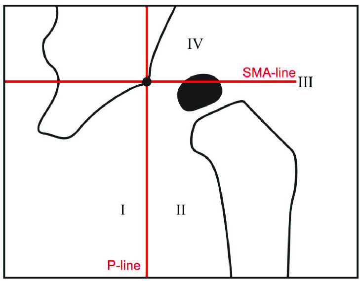
tonnis classification DDH
Terjesen, Terje & Horn, Joachim. (2020). Prognostic value of severity of dislocation in late-detected developmental dysplasia of the hip. Journal of Children’s Orthopaedics. 14. 1-7. 10.1302/1863-2548.14.200079. [CC BY SA 4.0]
  • Grade I: Ossific nucleus is medial to P-line (Infero-medial quadrant)
  • Grade II: Ossific nucleus is lateral to P-line but inferior to SMA-line (Infero-lateral quadrant)
  • Grade III: Ossific nucleus is lateral to P-line leveled to SMA-line
  • Grade IV: Ossific nucleus in superior to SMA-line (Supero-lateral quadrant)

IHDI classification

The position of the proximal femoral metaphysis (instead of the ossific nucleus) is used as the important reference landmark to determine the position of the femoral head. This classification requires supine AP X-ray of pelvis with the hips at rest while the lower limbs are gently held in neutral position without traction and patellae forward.

  • H-point is the midpoint of the superior margin of the ossified metaphysis
  • H-line is drawn through the top of the tri-radiate cartilages bilaterally
  • P-line is then drawn perpendicular to the H-line at the superolateral margin of the acetabulum
  • An additional diagonal line (D-line) is then drawn 45 degrees from the junction of H-line and P-line
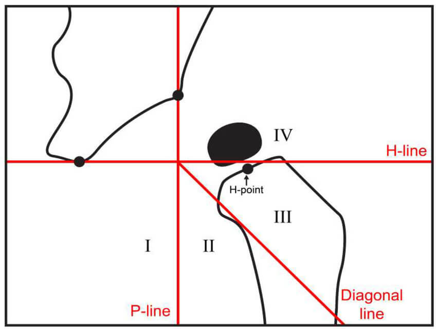
IHDI classification ddh
Terjesen, Terje & Horn, Joachim. (2020). Prognostic value of severity of dislocation in late-detected developmental dysplasia of the hip. Journal of Children’s Orthopaedics. 14. 1-7. 10.1302/1863-2548.14.200079. [CC BY SA 4.0]

The position of the H-point then determines the IHDI grade:

  • IHDI grade I: H-point is at or medial to the P-line
  • IHDI grade II: H-point is lateral to the P-line and at or medial to the D-Line
  • IHDI grade III: H-point is lateral to the D-line and at or inferior to the H-line
  • IHDI grade IV: H-point is superior to the H-line

References:

1. IHDI Standards – International Hip Dysplasia Institute

2. Pasupathy B, Sathish M. Reliability of a New Radiographic Classification System for Developmental Dysplasia of the Hip. International Journal of Paediatric Orthopaedics Jan-April 2020; 6(1): 16-19.

dr. sulabh kumar shrestha
Dr. Sulabh Kumar Shrestha, MS Orthopedics

He is the section editor of Orthopedics in Epomedicine. He searches for and share simpler ways to make complicated medical topics simple. He also loves writing poetry, listening and playing music. He is currently pursuing Fellowship in Hip, Pelvi-acetabulum and Arthroplasty at B&B Hospital.

  • Facebook
  • Twitter
PGMEE, MRCS, USMLE, MBBS, MD/MS OrthopedicsPediatric hipPediatrics

Post navigation

Previous post
Next post

Related Posts

PGMEE, MRCS, USMLE, MBBS, MD/MS open-closed angle glaucoma

Glaucoma basics : Classification of Glacuoma

Jun 4, 2014

Definition: Glaucoma is a group of disorders characterized by a progressive optic neuropathy resulting in a characterstic appearance of the optic disc and a specific pattern of irreversible visual field defects that are associated frequently but not invariably with raised intraocular pressure (IOP). It is one of the commonest cause of irreversible blindness in the world. Open-angle…

Read More
PGMEE, MRCS, USMLE, MBBS, MD/MS sacral plexus mnemonic

Lumbosacral Plexus Simplified

Aug 18, 2016Oct 17, 2020

While everyone is busy talking about the brachial plexus – lumbosacral plexus (the origin of nerves that supplies everything below the umbilicus) seems to be bit under-rated. Formation of Lumbosacral Plexus Ventral rami of L1-S4; has 2 components – Lumbar plexus (L1-L4) – forms within psoas major anterior to lumbar…

Read More
PGMEE, MRCS, USMLE, MBBS, MD/MS

Flow-Volume Loop : Mnemonics

Jun 19, 2024

The flow-volume loop is a plot of inspiratory and expiratory flow (on the Y-axis) against volume (on the X-axis) during the performance of maximally forced inspiratory and expiratory maneuvers. Condition Mnemonic Flow-volume loop Features Obstructive lung disease (small airways) Shift to leftConcave/Scooped out expiratory flow (↓FEV 25-75%) ↑TLC and RV↓FEV1…

Read More

Leave a Reply Cancel reply

Your email address will not be published. Required fields are marked *

This site uses Akismet to reduce spam. Learn how your comment data is processed.

Shrestha SK. Tonnis and IHDI (International Hip Dysplasia Institute) Classification for DDH [Internet]. Epomedicine; 2025 Nov 10 [cited 2026 Aug 20]. Available from: https://epomedicine.com/medical-students/tonnis-and-ihdi-international-hip-dysplasia-institute-classification-for-ddh/.

Pre-clinical (Basic Sciences)

Anatomy

Biochemistry

Community medicine (PSM)

Embryology

Microbiology

Pathology

Pharmacology

Physiology

Clinical Sciences

Anesthesia

Dermatology

Emergency medicine

Forensic

Internal medicine

Gynecology & Obstetrics

Oncology

Ophthalmology

Orthopedics

Otorhinolaryngology (ENT)

Pediatrics

Psychiatry

Radiology

Surgery

RSS feed: Ask Epomedicine Ask Epomedicine

  • What to study for Clinical examination in Orthopedics?
  • What is the mechanism of AVNRT?

Epomedicine weekly

  • About Epomedicine
  • Contact Us
  • Author Guidelines
  • Submit Article
  • Editorial Board
  • USMLE
  • MRCS
  • Thesis
©2026 Epomedicine | WordPress Theme by SuperbThemes