Skip to content
Epomedicine

Mnemonics, Simplified Concepts & Thoughts

Epomedicine

Mnemonics, Simplified Concepts & Thoughts

Popliteal Fossa : Mnemonics

Epomedicine, May 13, 2023Aug 9, 2023

Mnemonic: Serve And Volley Next Ball

  1. Semimembranosus/Semitendinosus
  2. Artery – Popliteal artery
  3. Vein – Popliteal vein
  4. Nerve – Tibial nerve and Common peroneal nerve
  5. Biceps femoris

Think of popliteal fossa as a diamond-shaped space. The floor is formed by Popliteus muscle.

popliteal fossa schematic

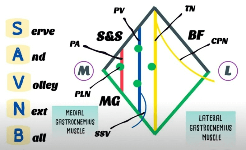
Membranosus = Medial (The 2 “Semis” go together – hence medial border is formed by semimembranosus, semitendinosus and medial head of gastrocnemius)

Therefore, biceps femoris and lateral head of gastrocnemius has to be lateral.

The tibial nerve lies superior to the vessels in the inferior aspect of the popliteal fossa. In the upper part of the fossa, the tibial nerve lies lateral to the vessels, it then passes superficial to them to lie medially. The popliteal artery is the deepest structure in the popliteal fossa.

Causes of lump/swelling in popliteal fossa

Mnemonic: ABCD

  1. Aneurysm
  2. Benign and malignant neoplasms:
    • Rhabdomyosarcoma
    • Pigmented villonodular synovitis (PVNS)
    • Synovial hemangioma
  3. Cysts:
    • Baker’s cyst (Synovial cyst)
    • Ganglionic cyst
    • Meniscal cyst
  4. DVT
  • Facebook
  • Twitter
PGMEE, MRCS, USMLE, MBBS, MD/MS Anatomy

Post navigation

Previous post
Next post

Related Posts

PGMEE, MRCS, USMLE, MBBS, MD/MS

Pelvic Lymphatic Drainage

Jun 26, 2024Jun 26, 2024

1. Para-aortic (lumbar) nodes: Gonads (derive blood supply from kidneys) 2. Inferior mesenteric nodes: As of blood supply – to the structures derived from hindgut 3. Common iliac nodes: Receives external and internal iliac nodes; Drains into para-aortic nodes 4. Superficial inguinal nodes: Everything that can be touched with fingers…

Read More
PGMEE, MRCS, USMLE, MBBS, MD/MS maxillary artery

Maxillary Artery : Mnemonic

Oct 16, 2017Aug 23, 2023

Origin of Maxillary artery: Terminal branch of External Carotid Artery (ECA) Derived from: 1st Arch Divisions of Maxillary artery: 3 parts by lateral pterygoid Branches of Maxillary artery Remember: Branches from 1st part Branches from the 2nd part Mnemonic: They supply muscles of mastication which are also derivatives of the…

Read More
PGMEE, MRCS, USMLE, MBBS, MD/MS ulnar nerve course

Ulnar nerve Anatomy – Course and Innervation

Jun 7, 2018Jun 1, 2024

Origin: C(7), C8, T1 (medial cord of Brachial plexus) Course: Motor innervation: 1. Forearm: Flexor carpi ulnaris (weakness of ulnar deviation and flexion of wrist), Medial half of flexor digitorum profundus (branches near the elbow) 2. Hand: branches near wrist Sensory innervation: Palmar branch at forearm and Digital branch at wrist…

Read More

Leave a Reply Cancel reply

Your email address will not be published. Required fields are marked *

This site uses Akismet to reduce spam. Learn how your comment data is processed.

Pre-clinical (Basic Sciences)

Anatomy

Biochemistry

Community medicine (PSM)

Embryology

Microbiology

Pathology

Pharmacology

Physiology

Clinical Sciences

Anesthesia

Dermatology

Emergency medicine

Forensic

Internal medicine

Gynecology & Obstetrics

Oncology

Ophthalmology

Orthopedics

Otorhinolaryngology (ENT)

Pediatrics

Psychiatry

Radiology

Surgery

RSS Ask Epomedicine

  • What to study for Clinical examination in Orthopedics?
  • What is the mechanism of AVNRT?

Epomedicine weekly

  • About Epomedicine
  • Contact Us
  • Author Guidelines
  • Submit Article
  • Editorial Board
  • USMLE
  • MRCS
  • Thesis
©2026 Epomedicine | WordPress Theme by SuperbThemes