Origin
L5-S1 (common iliac artery bifurcation; anterior to SI joint)
Course
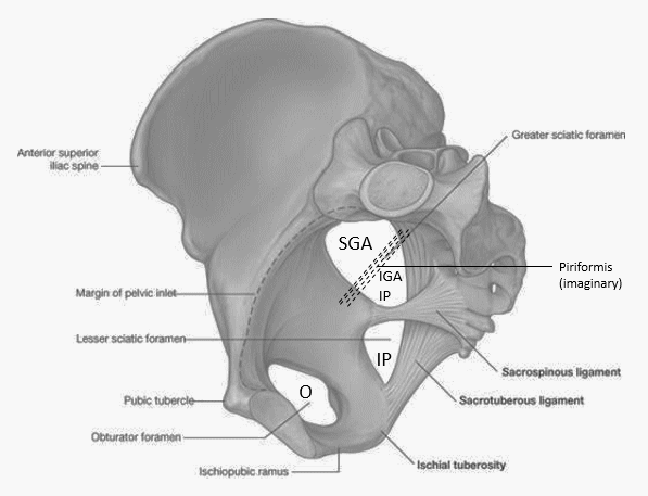
Extends down and posteriorly ~4 cm until superior margin of greater sciatic foramen and bifurcates into 2 trunks (in 60% cases) –
- Anterior trunk: continuation of the main artery towards ischial spine
- Posterior trunk: passes towards greater sciatic foramen

The anatomy is highly variable and may differ from books to books, person to person and even from right side to the left side in the same person.
Branches from anterior trunk
Four to pelvic viscera:

- Urinary bladder: Superior vesical artery, Inferior vesical artery (male)/Vaginal artery, Umbilical artery (in fetus; medial umbilical ligament in adults)
- Uterus/Vagin: Uterine artery, Vaginal artery (females)
- Rectum: Middle rectal artery
Three exits:

- Obturator artery: Exits pelvis through obturator foramen
- Internal pudendal artery: Exits pelvis through infra-piriform foramen
- Inferior gluteal artery: Exits pelvis through infra-piriform foramen
Branches from posterior trunk:
Mnemonic: PILS (Posterior trunk, Iliolumbar, Lateral sacral, Superior gluteal)
- Iliolumbar artery: runs anterolaterally towards the medial border of the psoas major, where it divides into the lumbar and iliac branches.
- Lateral sacral artery: passes lateral to anterior sacral foramina
- Superior gluteal artery: exits pelvis through supra-piriform foramen
Corona mortis: Corona mortis is an anastomotic branch between the inferior epigastric (from external iliac) and obturator (from internal iliac) vessels. It is located behind the superior pubic ramus at a variable distance from the symphysis pubis (range 40-96 mm). Compression of the internal iliac alone will not completely stop the bleeding from this anomalous artery since it has a contribution from the external iliac artery.
Pelvic fractures: Fractures of both the pelvis and acetabulum may be associated with injury to the major pelvic vessels (primary injury or clot dislodgement or iatrogenic trauma). The sources of bleeding have been reported to be more posterior (internal iliac vessels or their posterior branches) in patients with unstable posterior pelvic fractures. In lateral compression fractures, commonly anterior vessels are injured (pudendal or obturator vessels).


