Anterior Forearm (Compartment) Muscles
Total muscles: 8 (4 superficial + 1 intermediate + 3 deep)
Mnemonic: Do it yourself as shown in the figure below! Place your thenar/hypothenar eminence over medial epicondyle and fan out 5 fingers with thumb resting below the 4 fingers. The 4 fingers represent superficial flexors while the thumb represents intermediate/middle flexor. Starting from the index finger count – “Pass, Fail, Pass, Fail, and Fail (at last)” i.e. Pronator teres (PT), Flexor carpi radialis (FCR), Palmaris longus (PL), Flexor carpi ulnaris (FCU) and Flexor digitorum superficialis (FDS) resectively.
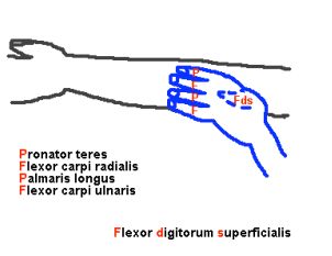
Superficial and Middle flexors
Common flexor origin: Medial epicondyle
But remember that the 3 muscles containing “U” have more attachments:
- Ulnar (deep) head of pronator teres (may be absent): Ulna (coronoid process)
- Flexor Carpi Ulnaris: Ulna (olecranon) and Ulnar shaft
- Flexor digitorum sUperficialis: Ulna (coronoid) and Radial shaft
Nerve supply: Median nerve except FCU which is supplied by ulnar nerve
| Muscle | Insertion | Function | Clinical relevance |
| Pronator teres (PT) | Mid-lateral radius | Pronate and Flex forearm | Median nerve compression (pronator syndrome) |
| Flexor carpi raialis (FCR) | 2nd and 3rd metacarpal base | Flex wrist, radial deviation | Radial artery lies between FCR and Brachioradialis |
| Palmaris longus (PL) | Flexor retinaculum/ Palmar aponeurosis | Flex wrist | Used for tendon transfers Congenitally absent in 10% |
| Flexor carpi ulnaris (FCU) | Pisiform (sesamoid – developed within tendon of FCU), hook of hamate, 5th metacarpal | Flex wrist, ulnar deviation | Most powerful flexor of wrist May compress ulnar nerve between 2 heads |
| Flexor digitorum superficialis (Sublimis; FDS) | Base of middle phalanges (at proximal phalanx splits into medial and lateral slips) | Flex Proximal Interphalangeal (PIP) joint | Sublimis test will isolate and test function |
Insertion of FCR on 2nd and 3rd metacarpal base matches the insertion of ECRL and ECRB.
Deep flexors
Mnemonic: All 3 deep flexors have a word starting with “P“.
1. flexor digitorum Profundus
2. flexor Pollicis longus
3. Pronator quadratus
All these 3 are supplied by AIN and can be tested with “OK” sign.
Nerve supply: Anterior interosseous branch of median nerve (AIN) except Flexor digitorum profondus of ring and little fingers which is also supplied by ulnar nerve.
Remember few things:
- FPL is for thumb, hence originates at the side of thumb (radius and interosseous membrane)
- FDS is for digits, hence originates at the side of the digits (ulna and interosseous membrane)
- Pronators, be it “teres” or “quadratus” goes from ulna to radius.
- “Longus” goes to distal phalanx base.
- “Superficialis” goes to proximal phalanx base.
| Muscle | Origin | Insertion | Function | Clinical relevance |
| Flexor Digitorum Profundus (FDP) | Anterior ulna and interosseous membrane | Base of distal phalanges (at proximal phalanx, it passes between 2 slips of FDS – camper’s chiasm) | Flex Distal Interphalangeal Joint (DIP) | Avulsion: Jersey finger Profundus test will isolate and test function |
| Flexor Pollicis Longus (FPL) | Anterior radius and interosseous membrane | Base of distal phalange of thumb | Flex Interphalangeal joint of thumb | FDP and FPL are most susceptible to Volkmann’s ischemic contracture (VIC) |
| Pronator Quadratus | Medial distal ulna (oblique ridge) | Volar distal radius | Pronate forearm | Primary pronator (initiates pronation) |
Remember the Rule of three (3):
3 wrist flexors: FCR, PL, FCU
3 finger flexors: FDS, FDP, FPL
Other 2 are pronators: PT, PQ
Posterior Forearm (Compartment) Muscles
Total muscles: 12 (4 Superficial + 3 Mobile wad + 5 Deep)
Superficial Externsors
Mnemonic: Do it yourself as shown in the picture! Hold your elbow with thumbs up and other 4 fingers curling behind the lateral epicondyle. Then go in the sequence of thumb (B-C), little finger (C-D), ring finger (D), middle finger (C) and index finger (A). The thumb is above and represents the origin point as lateral condyle for Brachioradialis and extensor Carpi radialis longus (B-C). All other fingers represent origin point as lateral epicondyle: Little finger – extensor Carpi radialis brevis, extensor Digitorum (C-D); Ring finger – extensor Digitorum minimi (D); Middle finger – extensor Carpi ulnaris; Index finger – Anconeus (A).
Common extensor origin: Lateral epicondyle
- Brachioradialis and ECRL originates from: Lateral supracondylar ridge
Nerve supply: Radial nerve or one of it’s branches
- ECRB, EDC, EDM, ECU (lateral epicondyle group) are supplied by raidal nerve branche – PIN (posterior interosseous nerve).
Remember:
- ECU is a large muscle like FCU and has origin on posterior ulna too.
- ECRL and ECRB like FCR insert into 2nd and 3rd metacarpal base respectively.
- ECU like FCU inserts on 5th metacarpal base.
| Muscle | Insertion | Function | Clinical relevance |
| Brachioradialis (BR) | Lateral distal radius/styloid | Flex forearm | Deforming force in distal radius fracture; Radial artery lies medially |
| Extensor carpi radialis longus (ECRL) | 2nd Metacarpal base | Extend wrist | |
| Extensor carpi radialis brevis (ECRB) | 3rd Metacarpal base | Extend wrist | Degenerates in tennis elbow |
| Anconeus | Proximal dorsal ulna | Extend forearm | Muscular plane in Kocher approach |
| Extensor digitorum (EDC) | MCP (Metacarpophalangeal joint): Saggital band P2 (middle phalanx): Central slip P3 (distal phalanx): Lateral slip unite | Extend digits | Tendon avulsion: P2 – Boutonniere P3 – Mallet finger |
| Extensor digitorum minimi – 2 tendons (EDM/EDQ) | As above but in little finger | Extend little finger | Medial to EDC to little finger |
| Extensor carpi ulnaris (ECU) | 5th Metacarapal base | Extend/adduct hand | Can cause painful snapping over ulna |
Brachioradiailis, ECRL and ECRB are the components of Mobile wad of Henry. These structures can be grasped between thumb and index finger just distal to lateral epicondyle. Brachioradialis is a paradoxical muscle. Its origin and innervation are characteristic of a extensor muscle, but it is actually a strong flexor of forearm.

Deep Extensors
Mnemonic: Like the deep flexor muscles, all the extensor muscles that have “P” in them are deep extensors –
1. abductor Pollicis longus (APL)
2. extensor Pollicis brevis (EPB)
3. extensor Pollicis longus (EPL)
4. extensor indicis Proprius (EIP)
5. PIN – supply all these muscles
6. SuPinator
Nerve supply: Radial nerve – PIN
Remember few things:
- Supinator like Pronator teres has also origin from the epicondyle (lateral epicondyle in contrast to medial epicondyle in pronator teres)
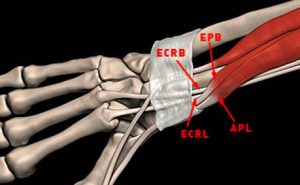
- Remember the 6 extensor compartments of wrist. The 1st compartment muscle (APL and EPB), they originate from radius and interosseous membrane.
- The 3nd and 4th compartment muscle (EPL and EIP respectively) originate from ulna and interosseous membrane.
| Muscle | Origin | Insertion | Function | Clinical relevance |
| Supinator | Posterior medial ulna including supinator crest | Proximal lateral radius | Supinate forearm | PIN can be compressed as it pierces the muscle |
| Abductor Pollicis Longus (APL) | Dorsal ulna and radius | 1st Metacarpal base | Abduct and extend thumb (CMCJ) | deQuervain’s tenosynovitis (may have multiple slips) |
| Extensor Pollicis Brevis (EPB) | Dorsal radius and interosseous membrane (below APL) | Base of proximal phalanx of thumb | Metacarpo-phalangeal (MCP) joint extension of thumb | Radial border of snuffbox |
| Extensor Pollicis Longus (EPL) | Posterior ulna and interosseous membrane (below APL) | Base of distal phalanx of thumb | Interphalangeal joint (IPJ) extension of thumb | Tendon turns 45 degrees on Lister’s tubercle |
| Extensor Indicis Proprius (EIP) | Posterior ulna and interosseous membrane (below EPL) | Same as EDC | Extension of index finger | Ulnar to EDC tendon; last PIN muscle |
From superior to inferior, origin of muscles are:
a. APL (radius and ulna) – remember A for Above
b. EPL (ulna) and EPB (radius)
d. EIP (ulna)
As in flexors, we have rule of threes (3) for extensors as well:
3 muscles for wrist extension: ECRB, ECRL, ECU
3 muscles for finger extension: EDC, EIP, EDM
3 muscles for thumb: EPL, EPB, APL
Another handy relation to keep in the back of head is: longus, brevis, longus, brevis (longus is lateral to brevis)
In the distal forearm, APL and EBP crosses from medial to lateral over ECRL and ECRB. APL and EPB enter 1st extensor compartment at wrist while the ECRL and ECRB enter the 2nd.
Recommended reading: Netter’s atlas for images

He is the section editor of Orthopedics in Epomedicine. He searches for and share simpler ways to make complicated medical topics simple. He also loves writing poetry, listening and playing music. He is currently pursuing Fellowship in Hip, Pelvi-acetabulum and Arthroplasty at B&B Hospital.
Thanks alot for the tricks. Made it alot easier to memorize them.
This is so awesome, thank you!
Very relevant and precise easy to remember information. Thanks
Thank you so much. Will try this!!
Thank you very much Sir. This helped me in my professional exams
thank you