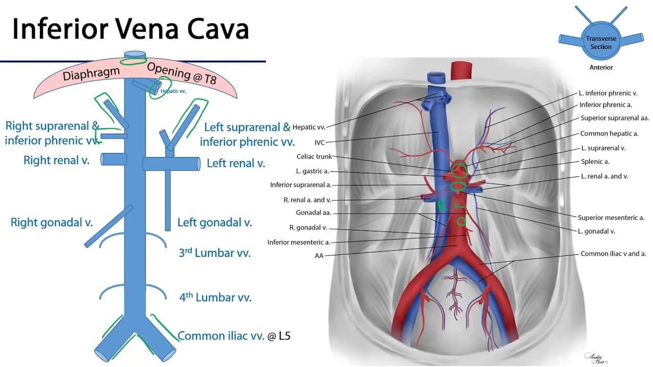
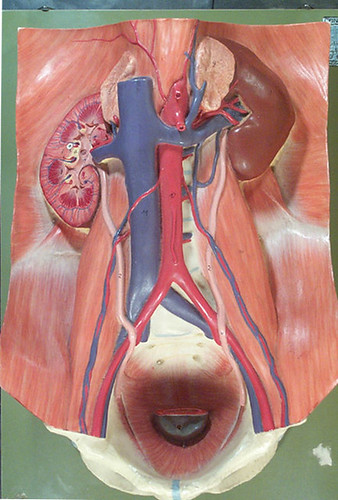
Inferior Venacava (IVC) Tributaries
Mnemonic: I Like To Rise So Incredibly High
1. common Iliac veins (L5) – Origin or Formation
- Median sacral vein ends in left common iliac vein or the junction of 2 common iliac veins
2. Lumbar veins (L1-L5)
3. Testicular/gonadal veins – right (L2)
4. Renal veins (L1)
- Since the IVC is not a midline structure, there is a degree of asymmetry of drainage, e.g. the gonadal and suprarenal veins drain into the IVC on the right side, but into the left renal vein on the left.
5. Suprarenal veins – right (L1)
6. Inferior phrenic veins (T8)
7. Hepatic veins (T8)
Abdominal Aorta Branches
Mnemonic: Cesarean Section In Some Races Induces Irregular Late Menstrual Cycle
| Branches | Group | Level |
| Celiac artery | Anterior visceral | T12 |
| Superior mesenteric artery | “ | L1 |
| Inferior mesenteric artery | “ | L3 |
| Suprarenal artery – middle (1 pair) | Lateral visceral | T12 |
| Renal artery (1 pair) | “ | L1 |
| Internal spermatic or ovarian artery (1 pair) | “ | L2 |
| Inferior phrenic artery (1 pair) | Lateral abdominal | T12 |
| Lumbar artery (4 paired) | Lateral abdominal/Dorsal | L1-L4 |
| Median sacral artery | Terminal/Dorsal | L4 |
| Common iliac arteries (1 pair) | Terminal | L4 |
Superior suprarenal artery originates from Inferior phrenic artery.
Inferior suprarenal artery originates from Renal artery.
Right renal artery passes behind the IVC.
Relationship of abdominal aorta with inferior vena cava
Abdominal aorta travels on left.
IVC travels on right side parallelly, and both can be considered counterparts.
Above the level of the umbilicus:
- Aorta is posterior to IVC:
- Right renal artery (longer) travels behind IVC.
- Left renal vein (longer) travels anterior to aorta.
Below the level of the umbilicus:
- Aorta is anterior to IVC:
- Common iliac arteries are anterior and lateral in relation to common iliac veins.
- Right common iliac artery (longer) travels anterior to left common iliac vein (longer)

Tajiri applying camel clutch to Rene Bonaparte. Remember this picture as a mnemonic for the relation between IVC and aorta. Tajiri can be likened to aorta and Rene can be likened to IVC.

He is the section editor of Orthopedics in Epomedicine. He searches for and share simpler ways to make complicated medical topics simple. He also loves writing poetry, listening and playing music. He is currently pursuing Fellowship in Hip, Pelvi-acetabulum and Arthroplasty at B&B Hospital.