Skip to content
Epomedicine

Mnemonics, Simplified Concepts & Thoughts

Epomedicine

Mnemonics, Simplified Concepts & Thoughts

Liver Anatomy : Mnemonics

Epomedicine, May 13, 2024May 13, 2024

Ligaments

Mnemonic: TV TFCL

1. True ligaments:

  • ligamentum Teres hepatis/round ligament (obliterated umbilical vein remnant)
  • ligamentum Venosum (ductus venosus remnant)

2. False ligaments (Peritoneal folds):

  • Triangular ligaments (right and left): connects respective liver lobes to diaphragm
  • Falciform ligament: connects anterior surface of liver to anterior abdominal wall
    • it’s free edge contains ligamentum teres
  • Coronary ligament: encloses bare area of liver
    • anterior and posterior folds on each side unites to form triangular ligament
  • Lesser omentum:
    • Hepatogastric ligament
    • Hepatoduodenal ligament (surrounds portal triad)

Posteroinferior (Visceral) Surface of Liver
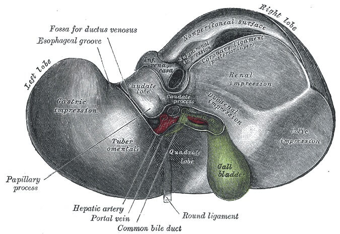
posteroinferior surface liver
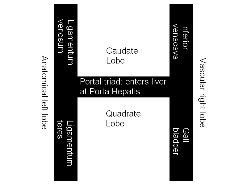
schematic posteroinferior liver surface

Anatomical halves are separated by Ligamentum venosum and Ligamentum teres.

Vascular halves are separated by IVC and Gall bladder.

Caudate lobe and Quadrate lobe:

  • Anatomically: Right lobe
  • Functionally (Vascular): Left lobe

Mnemonic: IVC are in one line

  • Inferior venacava (IVC)
  • ligamentum Venosum
  • Caudate lobe

Mnemonic: LGBTQ are in one line

  • Ligamentum Teres
  • Gall Bladder
  • Quadrate lobe

Porta hepatis separates the caudate and quadrate lobes. It transmits all the vessels, nerves and ducts entering or leaving the liver with the exception of the hepatic veins. The order of the structures in the porta hepatis from anterior to posterior can be remembered using the mnemonic “DAVE”:

  1. Ducts (right and left hepatic duct branches)
  2. Arteries (right and left hepatic artery branches)
  3. Vein (portal vein; NOT hepatic vein)
  4. Epiploic foramen (of Winlsow)

Hepatic Segmentation

Liver Segments Explained with Mnemonic

Liver Zonation

Mnemonic: Cen-three-lobular zone

Functionally, liver can be divided into 3 zones, based upon oxygen supply.

  1. Zone 1: Periportal zone
    • Predominantly involved in autoimmune and viral hepatitis (ALT > AST)
  2. Zone 2: Transitional zone
  3. Zone 3: Centrilobular zone
    • Predominantly involved in ischemic and toxic events, heart failure and Budd-chiari syndrome (AST > ALT)
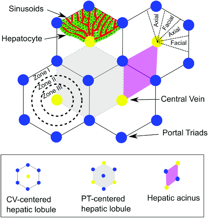
liver lobules

Blood and Nerve Supply of Liver

1. Arteries:

  • Hepatic artery (30%) – branch of celiac trunk
  • Portal vein (70%)

2. Veins: IVC via right and left hepatic veins

3. Lymphatics: Celiac nodes

4. Nerves:

  • Sympathetic: Celiac plexus
  • Parasympathetic: Anterior vagal trunk
  • Facebook
  • Twitter
PGMEE, MRCS, USMLE, MBBS, MD/MS AnatomyGastrointestinal system

Post navigation

Previous post
Next post

Related Posts

PGMEE, MRCS, USMLE, MBBS, MD/MS

Trichomycosis Axillaris : Malodorous yellowish armpit hair

Apr 24, 2021Apr 24, 2021

Synonym: Trichobateriosis axillaris, Bacterial trichonodosis, Trychomycosis palmellina Definition: Asymptomatic superficial bacterial infection (mycosis is a misnomer because it is not a fungal infection) primarily of the axillary hairs (can also occur in pubic, perianal and scalp hair) caused by Corynebacterium sp. Causative organism: Corynebacterium species particularly Corynebacterium flavescens Mode of infection: Bacteria…

Read More
PGMEE, MRCS, USMLE, MBBS, MD/MS parinaud syndrome

Parinaud Syndrome – Anatomical Basis

May 19, 2019May 19, 2019

Synonyms: Dorsal midbrain sydnrome, Pretectal syndrome Main components of Parinaud syndrome: Vertical gaze palsy Convergence-retraction nystagmus Pupillary light-near dissociation Lid retraction (Collier’s sign) Anatomical basis of Parinaud syndrome: Site of lesion: Dorsal midbrain Close to cerebral aqueduct and pineal gland – hydrocephalus and pinealoma Arterial supply – posterior cerebral artery…

Read More
PGMEE, MRCS, USMLE, MBBS, MD/MS Ostiomeatal unit anatomic variants

Anatomy of Ostiomeatal complex

May 14, 2014May 14, 2014

Synonyms: Ostiomeatal unit, Osteomeatal complex, OMC Definition: The term “ostiomeatal unit” represents the area on the lateral nasal wall (middle meatus) that receives drainage from the anterior  and medial ethmoid cells, frontal sinus, and maxillary sinus. It is an antomically constricted area that is prone to blockage, especially in the…

Read More

Leave a Reply Cancel reply

Your email address will not be published. Required fields are marked *

This site uses Akismet to reduce spam. Learn how your comment data is processed.

Epomedicine. Liver Anatomy : Mnemonics [Internet]. Epomedicine; 2024 May 13 [cited 2026 Jul 22]. Available from: https://epomedicine.com/medical-students/liver-anatomy-mnemonics/.

Pre-clinical (Basic Sciences)

Anatomy

Biochemistry

Community medicine (PSM)

Embryology

Microbiology

Pathology

Pharmacology

Physiology

Clinical Sciences

Anesthesia

Dermatology

Emergency medicine

Forensic

Internal medicine

Gynecology & Obstetrics

Oncology

Ophthalmology

Orthopedics

Otorhinolaryngology (ENT)

Pediatrics

Psychiatry

Radiology

Surgery

RSS Ask Epomedicine

  • What to study for Clinical examination in Orthopedics?
  • What is the mechanism of AVNRT?

Epomedicine weekly

  • About Epomedicine
  • Contact Us
  • Author Guidelines
  • Submit Article
  • Editorial Board
  • USMLE
  • MRCS
  • Thesis
©2026 Epomedicine | WordPress Theme by SuperbThemes