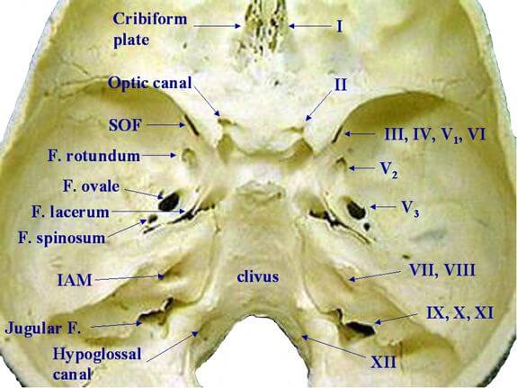
Skip to content
Epomedicine

Mnemonics, Simplified Concepts & Thoughts

Epomedicine

Mnemonics, Simplified Concepts & Thoughts

Middle Cranial Fossa : Mnemonics

Epomedicine, Jun 3, 2023Apr 29, 2024

Boundaries

  1. Anteriorly and Laterally: Greater and lesser wings of sphenoid
  2. Posteriorly: Clivus
  3. Posteromedially: Petrous part of temporal bone
  4. Floor: Greater wings of sphenoid and Petrous part of temporal bone
middle cranial fossa
Polygon data were generated by Database Center for Life Science (DBCLS), CC BY-SA 2.1 JP, via Wikimedia Commons.
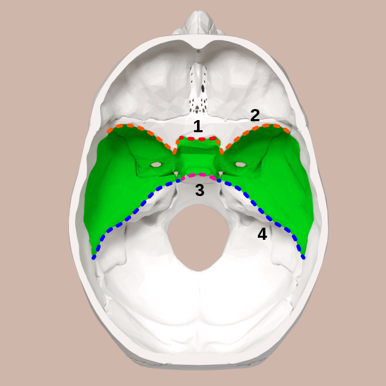
1. Sphenoidal limbus
2. Lesser wing of sphenoid
3. Dorsum sellae of sphenoid bone
4. Petrous part of temporal bone

The body of the sphenoid makes up the central portion of the middle fossa and houses the sella turcica, bounded by the anterior and posterior clinoid processes.

List of Foramina from Anterior to Posterior

Mnemonic: Old Rotund Owls Spin Lazily

owl spin mnemonic
  1. Orbital fissure (Superior orbital fissure, i.e. SOF)
  2. foramen Rotundum
  3. foramen Ovale
  4. foramen Spinosum
  5. foramen Lacerum

These foramina are in the Sphenoid bone.

cranial fossa foramina

Contents of Foramina

1. Superior orbital fissure:

Mnemonic: Everything related to Eyes (LR6 SO4 Rest 3 + CN V1)

  1. CN III (Occulomotor nerve)
  2. CN IV (Trochlear nerve)
  3. CN V1 (Ophthalmic nerve – Lacrimal, frontal and nasociliary branches)
  4. CN VI (Abducens nerve)
  5. Ophthalmic vein

2. Foramen rotundum:

Mnemonic: RoMax (CN V2)

  1. CN V2 (Maxillary nerve)

3. Foramen ovale:

Mnemonic: O-Man/O-MALE including CN V3

  1. Otic ganglion
  2. CN V3 (Mandibular nerve)
  3. Accessory meningeal artery
  4. Lesser petrosal nerve (parasympathetic fibers from glossopharyngeal nerve which receives a connecting branch from the facial nerve and then joins the otic ganglion)
  5. Emissary vein

4. Foramen spinosum:

Mnemonic: MMS

  1. Middle meningeal artery
  2. Meningeal branch of mandibular artery

5. Foramen lacerum/Carotid canal:

Mnemonic: LIP

  1. Internal carotid artery
  2. Pterygoid canal (Vidian canal) artery and nerve
    • Vidian artery: branch of maxillary artery
    • Vidian nerve: Greater superficial petrosal nerve + Deep petrosal nerve (ends in pterygopalatine ganglion)

Contents

  1. Temporal lobe
  2. Pituitary gland
  3. Trigeminal or Gasserian ganglion
  4. Greater superficial petrosal nerve (GSPN)
  5. Intracranial portion of ICA
  6. Cavernous sinus

Clinical considerations

Medial meningeal artery (MMA):

  • Ascends across the pterion (thin sheet of bone)
  • Auriculotemporal nerve wraps around it
  • Injury results in Extradural hemorrhage

Basilar skull fracture: can result to a carotid-cavernous fistula, manifesting with proptosis, chemosis, and pulsating exophthalmos

Vidian nerve: landmark for safe identification of petrous part of ICA

Greater petrosal nerve: reliable lateral landmark of Kawase triangle (laterally – GPN, anteriorly – CN V1, posteriorly – arcuate eminence)

1 shares
  • Facebook
  • Twitter
PGMEE, MRCS, USMLE, MBBS, MD/MS AnatomyNervous system

Post navigation

Previous post
Next post

Related Posts

PGMEE, MRCS, USMLE, MBBS, MD/MS

Arthrodesis Positions

Jul 26, 2022Jul 26, 2022

Arthrodesis is the surgical fusion of a diseased joint for the purposes of obtaining pain relief and stability. The bones are fused in a position permitting most useful function but lose their natural motion. Shoulder Flexion: 30° Abduction: 20° Internal rotation: 40° Elbow One side: 90° Both side: One in…

Read More
PGMEE, MRCS, USMLE, MBBS, MD/MS

Anterior Cranial Fossa : Mnemonic

Apr 29, 2024Apr 29, 2024

Boundaries Contents Mnemonic: Remember 6 ‘C’ 1. Frontal Crest: Site of attachment of falx cerebri 2. Foramen Cecum (Between frontal crest and crista galli): Site of communication between the draining veins of nasal cavity and the superior sagittal sinus 3. Crista galli: Site of attachment for falx cerebri 4. Cribiform…

Read More
PGMEE, MRCS, USMLE, MBBS, MD/MS fetal skull transverse

Fetal Skull Dimensions : Mnemonic

Jul 27, 2018

Anteroposterior Diameters Mnemonic: Larger the alphabets, larger the diameter (V>F>B) Diameters with “sub” prefix are smaller than counterpart without it. mento-Vertical: 14 cm SUB-mento-Vertical: 11.5 cm occipito-Frontal: 11.5 cm SUB-occipito-Frontal: 10.5 cm sub-occipito-Bregmatic: 9.5 cm sub-mento-Bregmatic: 9.5 cm Transverse Diameters Mnemonic: MTP (Medical Termination of Pregnancy) in increasing order. bi-Mastoid:…

Read More

Leave a Reply Cancel reply

Your email address will not be published. Required fields are marked *

This site uses Akismet to reduce spam. Learn how your comment data is processed.

Pre-clinical (Basic Sciences)

Anatomy

Biochemistry

Community medicine (PSM)

Embryology

Microbiology

Pathology

Pharmacology

Physiology

Clinical Sciences

Anesthesia

Dermatology

Emergency medicine

Forensic

Internal medicine

Gynecology & Obstetrics

Oncology

Ophthalmology

Orthopedics

Otorhinolaryngology (ENT)

Pediatrics

Psychiatry

Radiology

Surgery

RSS Ask Epomedicine

  • What to study for Clinical examination in Orthopedics?
  • What is the mechanism of AVNRT?

Epomedicine weekly

  • About Epomedicine
  • Contact Us
  • Author Guidelines
  • Submit Article
  • Editorial Board
  • USMLE
  • MRCS
  • Thesis
©2026 Epomedicine | WordPress Theme by SuperbThemes