Skip to content
Epomedicine

Mnemonics, Simplified Concepts & Thoughts

Epomedicine

Mnemonics, Simplified Concepts & Thoughts

Coronary Arteries : Mnemonic

Epomedicine, Jun 12, 2023Jun 12, 2023

Right coronary artery (RCA)

Course:

  1. Origin: Anterior aortic sinus (right cusp) of ascending aorta
  2. Between right auricle and pulmonary trunk root
  3. Right anterior coronary (atrioventricular) sulcus
  4. Winds round the inferior border of heart
  5. Right posterior coronary sulcus
  6. Posterior interventricular groove
  7. Anastomose with left coronary artery

Branches:

Mnemonic: TRaP Me IN

Large:

  • Marginal (largest branch)
    • Accompanied by Small cardiac vein
    • Supplies: Right ventricle
  • Posterior Interventricular (Posterior Descending Artery/PDA)
    • Accompanied by Middle cardiac vein
    • Supplies: Diaphragmatic surface of both ventricles, Septal branch of AVN, Posterior part of interventricular septum
    • Cardiac dominance: In 65% cases, PDA arise from RCA (right dominance) and in other cases from LCA (left dominance)
      • misleading term: because LCA always supplies greater myocardial portion

Small:

  • Nodal: Supplies SAN and both atria
  • Right atrial
  • Infundibular
  • Terminal
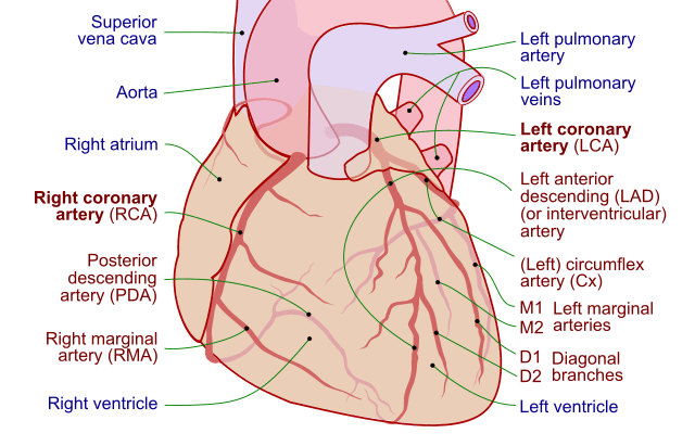
Coronary arteries
Coronary.pdf: Patrick J. Lynch, medical illustratorderivative work: Fred the Oysteradaption and further labeling: Mikael Häggström, M.D. Author info – Reusing images- Conflicts of interest:  NoneMikael Häggström, M.D., CC BY-SA 3.0, via Wikimedia Commons

Left coronary artery (LCA)

Course:

  1. Origin: Left posterior aortic sinus of ascending aorta
  2. Between pulmonary trunk and the left auricle
  3. Anterior interventricular groove
    • Anterior interventricular artery: to apex of heart
    • Circumflex artery (LCA): left anterior coronary sulcus → winds round left margin of heart → left posterior coronary sulcus → Supplies posterolateral wall of left ventricle
  4. Anastomose with right coronary artery

Branches:

Mnemonic: LaTe PAD

Large: Left Anterior Descending (LAD) artery –

  • Anterior Interventricular
    • Accompanied by Great cardiac vein
    • Supplies: Anterior 2/3 of interventricular septum
  • Diagonal branch
    • Supplies: Lateral wall of left ventricle

Small:

  • Left atrial
  • Pulmonary conus
  • Terminal

Arterial supply of conducting system of heart

1. RCA: SA node, AV node, AV bundle

2. LCA: Right bundle branch of AV bundle

3. Both RCA and LCA: Left bundle branch of AV bundle

Division of Coronary arteries

Coronary arteries → Epicardiac arteries → Final/intramural vessles
  • Facebook
  • Twitter
PGMEE, MRCS, USMLE, MBBS, MD/MS AnatomyCardiovascular system

Post navigation

Previous post
Next post

Related Posts

PGMEE, MRCS, USMLE, MBBS, MD/MS granuloma morphology pathophysiology

Morphology of Granuloma

Jan 19, 2017Jan 31, 2017

Concentric layers of Granuloma There are 4 concentric layers in a granuloma, however the clear distinction is difficult in reality due to overlapping. From inside to out: 1. Necrosis Caseating necrosis: Tuberculosis, Leprosy Coagulative necrosis: Buruli ulcer (M.ulcerans), Gumma containing central blood vessels (Syphilis) Fibrinoid necrosis: Aschoff bodies (Rheumatic granuloma),…

Read More
PGMEE, MRCS, USMLE, MBBS, MD/MS brachial plexus

Brachial Plexus Simplified with Mnemonics

Aug 19, 2016Dec 13, 2024

Components of Brachial Plexus Mnemonic: Randy Travis Drinks Cold Beer From proximal to distal, brachial plexus consists of: How are the roots formed? From the Ventral Rami of C5 to T1 spinal nerves. Extent and course: Intervertebral foramina to Transverse process to Interscalene triangle (bounded by anterior scalene and middle…

Read More
PGMEE, MRCS, USMLE, MBBS, MD/MS

Rheumatoid hand

Oct 2, 2022Oct 2, 2022

Ulnar drift or deviation of fingers Normal anatomical factors contributing to ulnar drift of fingers at MCP joint: Normal mechanical advantage of ulnar intrinsic muscles Asymmetry and ulnar slope of metacarpal heads of index and middle fingers Greater ulnar deviation permitted by radial collateral ligament when MCP joint is flexed…

Read More

Comment

  1. Neelam says:
    Jan 13, 2025 at 1:11 pm

    Thankyou for mnemonics of coronary artery cause as a average student it was hard for me to learn and I’m a perfusion student

    Reply

Leave a Reply Cancel reply

Your email address will not be published. Required fields are marked *

This site uses Akismet to reduce spam. Learn how your comment data is processed.

Epomedicine. Coronary Arteries : Mnemonic [Internet]. Epomedicine; 2023 Jun 12 [cited 2026 Sep 10]. Available from: https://epomedicine.com/medical-students/coronary-arteries-mnemonic/.

Pre-clinical (Basic Sciences)

Anatomy

Biochemistry

Community medicine (PSM)

Embryology

Microbiology

Pathology

Pharmacology

Physiology

Clinical Sciences

Anesthesia

Dermatology

Emergency medicine

Forensic

Internal medicine

Gynecology & Obstetrics

Oncology

Ophthalmology

Orthopedics

Otorhinolaryngology (ENT)

Pediatrics

Psychiatry

Radiology

Surgery

RSS feed: Ask Epomedicine Ask Epomedicine

  • What to study for Clinical examination in Orthopedics?
  • What is the mechanism of AVNRT?

Epomedicine weekly

  • About Epomedicine
  • Contact Us
  • Author Guidelines
  • Submit Article
  • Editorial Board
  • USMLE
  • MRCS
  • Thesis
©2026 Epomedicine | WordPress Theme by SuperbThemes