Skip to content
Epomedicine

Mnemonics, Simplified Concepts & Thoughts

Epomedicine

Mnemonics, Simplified Concepts & Thoughts

Coccyx Types

Epomedicine, May 23, 2025May 23, 2025

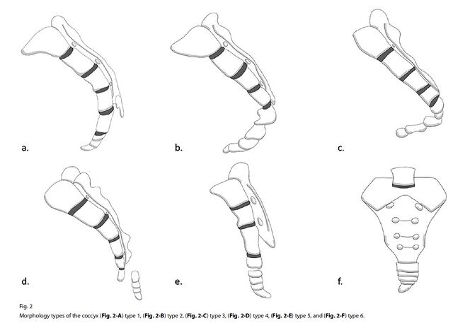
Postacchini and Massobrio Classification (Nathan Modification)

Can be classified into 6 types:

coccyx types
https://wikism.org/w/index.php?curid=4367

Type I (slight curve): Curved slightly forward with apex pointing caudally (>50% incidence)

Type II (forward curve): More prominent ventral curvature with coccyx apex pointing anteriorly (8-32% incidence)

Type III (sharp angle): Acute anterior angulation of coccyx but no subluxation (4-16% incidence)

Type IV (subluxation): Focal anterior angulation with anterior subluxation (1-9% incidence)

Type V (retroverted): Posteriorly angulated coccyx (1-11% incidence)

Type VI (scoliotic): Scoliotic deformity or lateral angulation of coccyx (1-6% incidence)

Clinical implication

Patients with a type II-IV coccyx are more prone to develop idiopathic coccygodynia

  • Facebook
  • Twitter
PGMEE, MRCS, USMLE, MBBS, MD/MS AnatomyMusculoskeletal systemOrthopedicsRadiology

Post navigation

Previous post
Next post

Related Posts

PGMEE, MRCS, USMLE, MBBS, MD/MS implantation

Embryology Week 1: Cleavage to Implantation

Aug 2, 2016Aug 2, 2016

Following events occur in the zygote formed in the week zero of development: Cleavage Approximately 24 hours after fertilization the zygote begins with the first cleavage division. Series of mitotic divisions of the zygote (occurs in fallopian tube) to form small daughter cells called blastomeres. Characteristics of cleavage in humans:…

Read More
PGMEE, MRCS, USMLE, MBBS, MD/MS

Well’s criteria for DVT with Mnemonic

Oct 9, 2020Jun 18, 2022

Mnemonic: ABCD(CD)E(CDE) – 4P A: Alternate diagnosis of DVT more likely (-2) B: Bedridden recently >3 days or Big (major) surgery within 3 months (+1) C: Cancer – treatment or palliation within 6 months (+1) D: Deep venous involvement: Mnemonic – CD Collateral (non-varicose) superficial veins present (+1) Deep venous…

Read More
PGMEE, MRCS, USMLE, MBBS, MD/MS Ostiomeatal unit anatomic variants

Anatomy of Ostiomeatal complex

May 14, 2014May 14, 2014

Synonyms: Ostiomeatal unit, Osteomeatal complex, OMC Definition: The term “ostiomeatal unit” represents the area on the lateral nasal wall (middle meatus) that receives drainage from the anterior  and medial ethmoid cells, frontal sinus, and maxillary sinus. It is an antomically constricted area that is prone to blockage, especially in the…

Read More

Leave a Reply Cancel reply

Your email address will not be published. Required fields are marked *

This site uses Akismet to reduce spam. Learn how your comment data is processed.

Pre-clinical (Basic Sciences)

Anatomy

Biochemistry

Community medicine (PSM)

Embryology

Microbiology

Pathology

Pharmacology

Physiology

Clinical Sciences

Anesthesia

Dermatology

Emergency medicine

Forensic

Internal medicine

Gynecology & Obstetrics

Oncology

Ophthalmology

Orthopedics

Otorhinolaryngology (ENT)

Pediatrics

Psychiatry

Radiology

Surgery

RSS Ask Epomedicine

  • What to study for Clinical examination in Orthopedics?
  • What is the mechanism of AVNRT?

Epomedicine weekly

  • About Epomedicine
  • Contact Us
  • Author Guidelines
  • Submit Article
  • Editorial Board
  • USMLE
  • MRCS
  • Thesis
©2026 Epomedicine | WordPress Theme by SuperbThemes