Skip to content
Epomedicine

Mnemonics, Simplified Concepts & Thoughts

Epomedicine

Mnemonics, Simplified Concepts & Thoughts

Ostiomeatal unit anatomic variants

Anatomy of Ostiomeatal complex

Epomedicine, May 14, 2014May 14, 2014

Synonyms: Ostiomeatal unit, Osteomeatal complex, OMC

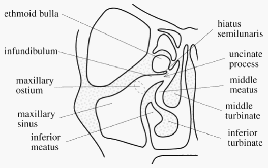
Definition: The term “ostiomeatal unit” represents the area on the lateral nasal wall (middle meatus) that receives drainage from the anterior  and medial ethmoid cells, frontal sinus, and maxillary sinus. It is an antomically constricted area that is prone to blockage, especially in the presence of structural anomalies, mucosal swelling or tumors. In addition, ostia themselves are small. An impairment in the ventilation of sinus due to such reasons lead to Chronic rhinosinusitis (CRS).

Boundaries:

  1. Medially: Middle turbinate
  2. Laterally: Lamina papyracea
  3. Superiorly and posteriorly: Basal lamella
  4. Inferiorly and anteriorly: It is open

Ostiomeatal unit

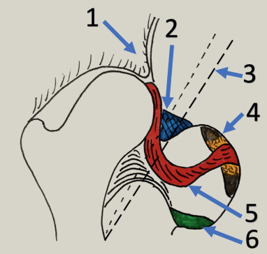
Components: Functionally, significant structures of the Ostiomeatal complex are:

  1. Uncinate process (Hook like bony extension of medial wall)
  2. Hiatus semilunaris (Crescent passage between uncinate process and ethmoid bulla through which middle meatus communicates with ethmoidal infundibulum)
  3. Frontal recess (Drainage channel of frontal sinus)
  4. Bulla ethmoidalis (most constant and largest anterior ethmoid air cell that projects inferomedially over hiatus semilunaris)
  5. Ethmoidal infundibulum (Funnel shaped passage through which anterior ethmoid cells and maxillary sinus drains into middle meatus)
  6. Maxillary sinus ostium (Drainage channel of maxillary sinus)
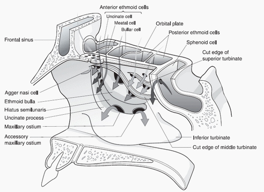
Lateral nasal wall

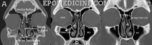
Anatomic variations:

  1. Deviated Nasal Septum
  2. Concha bullosa (Enlarged, pneumatized middle turbinate
  3. Intralamellar cell (Air cell witihin vertical portion of middle turbinate)
  4. Paradoxical middle concha (Convexity of turbinate directed towards lateral nasal wall)
  5. Haller cells (Infraorbital ethmoid air cells)
  6. Agger Nasi cells (Extension of anterior ethmoid air cells into lacrimal bone)
  7. Uncinate process bulla
  8. Deviation of uncinate process
Ostiomeatal unit anatomic variants
A: Concha bullosa
B: Haller cells
C: Agger Nasi cells

Silent sinus syndrome: Maxiallry sinus atelectasis due to chronic occlusion of maxillary sinus ostia, resulting in inward bowing of all 4 walls of the sinus including the orbital floor and increased orbital volume, leading to enophthalmos and hypoglobus (downward displacement of eye in orbit).

Note: According to some authors, basal lamella separates the anterior ostiomeatal complex (the one discussed above) and the posterior osteomeatal complex which is located in the spheno-ethmoidal recess and drains the posterior ethmoid and sphenoid sinuses. The posterior OMC is less involved in chronic sinusitis than the anterior one because its anatomic variations are fewer. So, when the disease is limited to the anterior OMC, basal lamella can be left undisturbed which reduces postoperative complications.
 

34 shares
  • Facebook34
  • Twitter
PGMEE, MRCS, USMLE, MBBS, MD/MS OtorhinolaryngologyRadiology

Post navigation

Previous post
Next post

Related Posts

PGMEE, MRCS, USMLE, MBBS, MD/MS eye schematic

Embryology of Eye : Mnemonic

Mar 3, 2019Mar 3, 2019

Surface ectoderm derivatives Mnemonic: LEVeL Lens Epithelium All ocular adnexa including mebomian gland and glands of Zeis and Moll Skin Cornea Conjunctiva Vitreous (portion) Lacrimal glands and drainage system Neuro-ectoderm (Optic vesicle and cup) derivatives Mnemonic: MORE Muscles of pupil (constrictor and dilator pupillae) Optic nerve Retinal pigment epithelium Epithelium…

Read More
PGMEE, MRCS, USMLE, MBBS, MD/MS fracture

Complications of Fractures

Oct 19, 2020Nov 4, 2022

This is a tabulated compilation for complications of fractures in general which can be immediate, early or delayed and local or generalized/systemic. This topic is commonly tested in exams. Local Systemic Immediate 1. Soft tissue injuries (Skin, Nerve, Vessels, Muscle-tendon) 2. Physeal injury 3. Hemarthrosis 4. Local visceral injury 1….

Read More
PGMEE, MRCS, USMLE, MBBS, MD/MS fetus

Non-invasive Prenatal Screening

Jun 8, 2019Mar 11, 2021

Quick review/Mnemonic: PAPP-A is decreased in all trisomies.Nuchal translucency is increased in all trisomies. Inhibin A is only increased in Down’s syndrome.Beta-HCG is increased in Down’s syndrome. All triple test results decreased in Edward syndrome. Only AFP is increased in NTDs and abdominal wall defects. Acetylcholinesterase level increased in Amniotic…

Read More

Comment

  1. Abdulwahab Alyahya says:
    May 17, 2020 at 3:03 pm

    Great information, thanks a lot..
    i’m currently working on a study that investigates the causes of performing revision frontal sinus surgery, may the authors of this article provide me with the references they used. thank you very much in advance.

    Abdulwahab Alyahya
    medical intern

    Reply

Leave a Reply Cancel reply

Your email address will not be published. Required fields are marked *

This site uses Akismet to reduce spam. Learn how your comment data is processed.

Epomedicine. Anatomy of Ostiomeatal complex [Internet]. Epomedicine; 2014 May 14 [cited 2026 Jul 5]. Available from: https://epomedicine.com/medical-students/anatomy-of-ostiomeatal-complex/.

Pre-clinical (Basic Sciences)

Anatomy

Biochemistry

Community medicine (PSM)

Embryology

Microbiology

Pathology

Pharmacology

Physiology

Clinical Sciences

Anesthesia

Dermatology

Emergency medicine

Forensic

Internal medicine

Gynecology & Obstetrics

Oncology

Ophthalmology

Orthopedics

Otorhinolaryngology (ENT)

Pediatrics

Psychiatry

Radiology

Surgery

RSS Ask Epomedicine

  • What to study for Clinical examination in Orthopedics?
  • What is the mechanism of AVNRT?

Epomedicine weekly

  • About Epomedicine
  • Contact Us
  • Author Guidelines
  • Submit Article
  • Editorial Board
  • USMLE
  • MRCS
  • Thesis
©2026 Epomedicine | WordPress Theme by SuperbThemes