Frontal Lobe
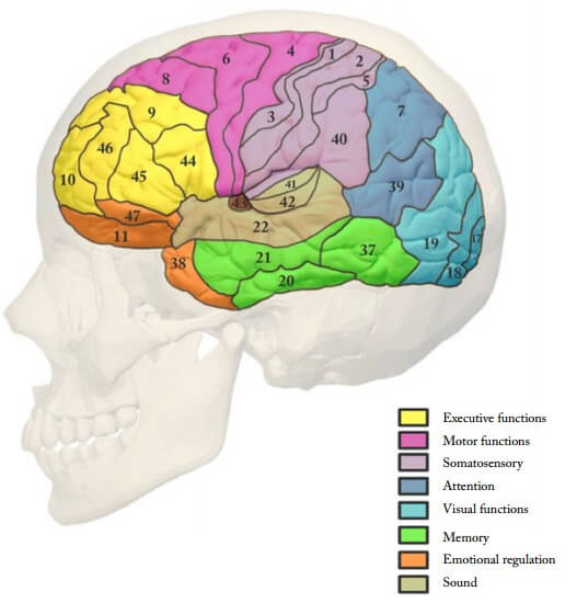
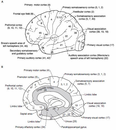
Area 4 (Precentral gyrus): Primary motor cortex (gigantopyramidal – only area that contains giant pyramidal cells of Betz)
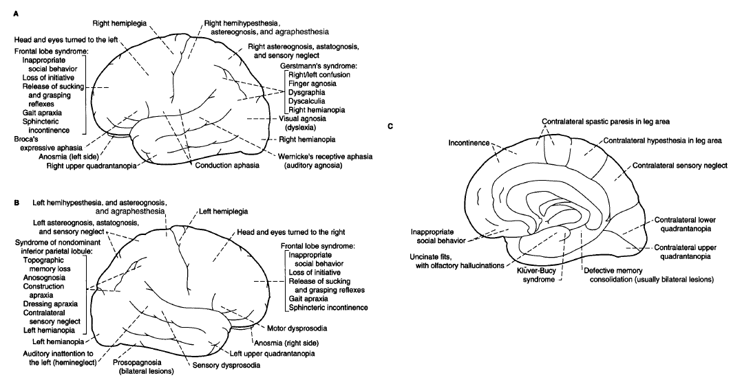
- Lesion: Contralateral spastic paralysis (UMNL)
Area 6 (Superior frontal gyrus; agranular frontal): Premotor cortex and Supplementary motor cortex (Motor planning)
- Lesion: Apraxia (Unable to perform movements in correct sequence)
Area 8 (Middle frontal gyrus; intermediate frontal): Frontal eye field (Contralateral horizontal conjugate eye movements)
- Lesion: Contralateral horizontal conjugate gaze palsy
Area 44 (Inferior frontal gyrus; pars opercularis) and Area 45 (Inferior frontal gyrus; pars triangularis): Broca’s area (Motor speech center only in Dominant hemisphere)
- Lesion: Comprehends language well but fails to express thoughts verbally or in written
- Nonfluent, motor or expressive aphasia
- Agraphia (inability to write)
Areas 9, 10, 46 (Prefrontal cortex) and Area 11, 47 (Orbitofrontal cortex): Part of limbic system regulating emotions and higher mental functions. Area 11 is associated with general olfaction.
- Lesion: Deficits in concentration, orientation, abstracting ability, judgement, problem-solving ability, loss of initiative, inappropriate behavior, frontal release of sucking and grasping reflexes.
Parietal Lobe
Areas 3,1 and 2 (Postcentral gyrus): Primary somatosensory cortex (Discriminative touch, vibration, position sense, pain and temperature)
- Lesion: Impairment of all somatic sensations in contralateral side of body.
Area 43 (Inferior parietal lobule, just below somatosensory cortex in postcentral gyrus): Primary gustatory area (sensory)
Areas 5 and 7 (Superior parietal lobule): Somatosensory association cortex (Spatial awareness and Awareness of body in general)
- Lesion: Contralateral astereognosis and sensory neglect (damage in nondominant hemisphere)
Area 40 (Inferior parietal lobule – Supramarginal gyrus) and Area 39 (Inferior parietal lobule – Angular gyrus): Multimodal association areas that receives input from visual, auditory and tactile modalities
These areas are also regarded as the part of Wernicke’s area along with area 22. Area 40 has strong connections with sensory areas and also regarded as somatosensory association area and Area 39 to the visual areas and also regarded as visual association cortex. Area 39 is also called “reading center” and also plays important role in arithmetic functions.
- Lesion in dominant hemisphere: Gerstmann syndrome
- Destruction of supramarginal gyrus (area 40): Disruption of connection to other sensory association cortices –
- Right and left confusion
- Destruction of angular gyrus (area 39): Disruption of connection to visual areas and arithmetic functions
- Finger agnosia (not a sensory agnosia but failure of recognition)
- Dysgraphia and dyslexia
- Dyscalculia
- Destruction of Baum’s loop: Contralateral hemianopia or lower quadrantanopia (pie in the floor).
- Destruction of supramarginal gyrus (area 40): Disruption of connection to other sensory association cortices –
- Lesion in non-dominant hemisphere:
- Topographic memory loss
- Anosognosia (lack of insight)
- Construction apraxia
- Dressing apraxia
- Contralateral sensory neglect
- Contralateral hemianopia or lower quadrantopia
Arcuate fasciculus: Connects Wernicke’s area with Broca’s area.
- Lesion: Conductive aphasia
- Comprehension is intact but repetition is poor and speech is fluent but paraphasic (word sounding the same as correct word but often making no sense)
Temporal lobe
Area 41 and 42 (Medial to Superior temporal gyrus – Transverse temporal gyrus of Heschl): Primary auditory cortex (Basic sound processing)
- Lesion: Unilateral lesion results in slight loss of hearing and Bilateral lesion results in cortical deafness.
Area 22 (Posterior part of Superior temproal gyrus): Wernicke’s area of sensory speech in dominant hemisphere (Auditory association cortex – Complex sound processing)
- Lesion: Sensory or fluent or receptive aphasia (failure to comprehend language but no problem in expression)
Area 21 (Middle temporal gyrus) and Area 20 (Inferior temproal gyrus): Part of auditory association cortex
Area 34 (Hippocampal – entorhinal area): Primary olfactory cortex
- Lesion: Ipsilateral anosmia
Area 28 (Hippocampal – uncal area):
- Uncal fits can lead to olfactory and gustatory hallucinations
Area 37 (Fusiform gyrus – occipitotemporal cortex): Familiar face recognition
- Lesion (Bilateral): Prosopagnosia (deficit for recognition of familiar faces, such as those of family, friends, and colleagues)
Occipital Lobe
Area 17 (striate cortex): Primary visual cortex
- Lesion: Contralateral homonymous hemianopia with macular sparing
Area 18 (peristriate cortex) and Area 19 (parastriate cortex): Visual association areas
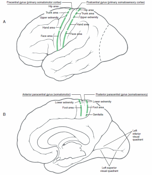
Motor and Sensory Homonculus or Somatotropy
The lower extremity and foot areas are located on medial aspects of the hemisphere in the anterior paracentral (motor) and the posterior paracentral (sensory) gyri.
The remaining portions of the body extend from the margin of the hemisphere over the convexity to the lateral sulcus in the precentral and postcentral gyri. An easy way to remember the somatotopy of these important cortical areas is to divide the precentral and postcentral gyri generally into thirds:
- Lateral 1/3rd: face area
- Middle 1/3rd: upper extremity and hand with particular emphasis on the hand
- Medial 1/3rd: trunk and hip
The mouth and hand, for instance, are disproportionately large because so much nervous system tissue is devoted to fine motor and sensory innervation.
I really appreciate work presented very clear and concise. Thanks