Skip to content
Epomedicine

Mnemonics, Simplified Concepts & Thoughts

Epomedicine

Mnemonics, Simplified Concepts & Thoughts

subclavian artery branches

Branches of Subclavian artery : Mnemonic

Epomedicine, Oct 10, 2017

Origin of Subclavian artery:

  • Left: Arch of aorta
  • Right: Brachiocephalic artery

Extent of Subclavian artery: Arises posterior to sternoclavicular joint and ends at outer border of 1st rib by becoming acillary artery.

Divisions of Subclavian artery: 3 parts in relation to Scalenus anterior muscle

  1. 1st part (medial to muscle)
  2. 2nd part (posterior to muscle)
  3. 3rd part (lateral to muscle)

subclavian artery branches

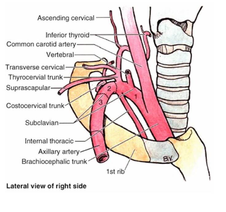
Branches of Subclavian artery:

Remember Vitamin C and D

Mnemonic: VIT(sit) C(sid) and D

  • Orange (VIT – from 1st part)
  • Green (C – from 2nd part)
  • Blue (D – from 3rd part)
  1. Vertebral artery
  2. Internal thoracic artery
  3. Thyrocervical trunk
    • Suprascapular artery
    • Inferior thyroid artery (Thyro-)
    • Transverse cervical artery (Cervical)
  4. Costocervical trunk
    • Superior intercostal (Costo-)
    • Deep cervical (Cervical)
  5. Dorsal scapular artery
172 shares
  • Facebook33
  • Twitter
PGMEE, MRCS, USMLE, MBBS, MD/MS AnatomyCardiovascular system

Post navigation

Previous post
Next post

Related Posts

PGMEE, MRCS, USMLE, MBBS, MD/MS

Oestern and Tscherne Classification for Closed fractures

Sep 6, 2020Sep 6, 2020

The classification system for closed fractures is based on the physiologic concept that the energy imparted to the bone (and the resultant fracture pattern) directly correlates with the energy transferred to the surrounding soft tissues. Grade Soft tissue injury Fracture Compartment C0 Absent or Negligible Simple (Spiral) Soft and/or Normal…

Read More
PGMEE, MRCS, USMLE, MBBS, MD/MS mathes and nahai

Mathes and Nahai Classification of Muscle Flap based on Vascular Anatomy

Oct 16, 2017

Type Dominant pedicle Minor pedicle Example I 1 – Tensor Fascia Lata (TFL) II 1 1 Gracilis III 2 – Gluteus maximus IV – Multiple Sartorius V 1 Multiple Lattisimus dorsi Mnemonic: Ten Graceful Glutes Sat on Latrines Type I: TFL Type II: Gracilis Type III: Gluteus maximus Type IV:…

Read More
PGMEE, MRCS, USMLE, MBBS, MD/MS

Flow-Volume Loop : Mnemonics

Jun 19, 2024

The flow-volume loop is a plot of inspiratory and expiratory flow (on the Y-axis) against volume (on the X-axis) during the performance of maximally forced inspiratory and expiratory maneuvers. Condition Mnemonic Flow-volume loop Features Obstructive lung disease (small airways) Shift to leftConcave/Scooped out expiratory flow (↓FEV 25-75%) ↑TLC and RV↓FEV1…

Read More

Comments (2)

  1. OBE BWAMI says:
    Oct 14, 2019 at 5:35 pm

    Well done

    Reply
  2. Farha says:
    Sep 20, 2023 at 2:54 am

    Very useful

    Reply

Leave a Reply to OBE BWAMI Cancel reply

Your email address will not be published. Required fields are marked *

This site uses Akismet to reduce spam. Learn how your comment data is processed.

Pre-clinical (Basic Sciences)

Anatomy

Biochemistry

Community medicine (PSM)

Embryology

Microbiology

Pathology

Pharmacology

Physiology

Clinical Sciences

Anesthesia

Dermatology

Emergency medicine

Forensic

Internal medicine

Gynecology & Obstetrics

Oncology

Ophthalmology

Orthopedics

Otorhinolaryngology (ENT)

Pediatrics

Psychiatry

Radiology

Surgery

RSS Ask Epomedicine

  • What to study for Clinical examination in Orthopedics?
  • What is the mechanism of AVNRT?

Epomedicine weekly

  • About Epomedicine
  • Contact Us
  • Author Guidelines
  • Submit Article
  • Editorial Board
  • USMLE
  • MRCS
  • Thesis
©2026 Epomedicine | WordPress Theme by SuperbThemes