Skip to content
Epomedicine

Mnemonics, Simplified Concepts & Thoughts

Epomedicine

Mnemonics, Simplified Concepts & Thoughts

Basics of voiding cystourethrogram (VCUG)

Dr. Manoj Bhandari, MS General Surgery Resident, Jan 12, 2024Jan 12, 2024
vcug basics
5 – penile urethra; 4 – bulbar urethra; 3 – bulbomembranous junction; 2 – verumontanum; 1 – prostatic urethra

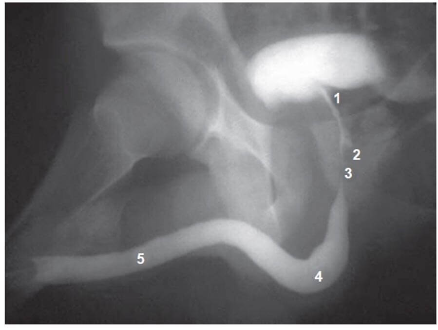
The above picture shows a VCUG scan which is a commonly performed test in urology for diagnosing various conditions related to urethra, bladder as well as assessing upper tracts. Diseases that can be diagnosed using VCUG includes:

  1. Urethral strictures and stenosis 
  2. Posterior urethral valves 
  3. Vesicourethral reflux 
  4. Uretroceles and many more 

So, it is always better to have an idea about interpreting the scan as a surgeon rather than relying on radiologists as they may not be available every time. 

This article will try to enumerate normal anatomical landmarks of urethra on the scan using above mentioned picture.

The male urethra has been divided into 2 main categories: 

  1. Anterior urethra (penile and bulbar urethra) 
  2. Posterior urethra (membranous and prostatic) 

In the picture above: 

5 – represents penile urethra which extends from meatus to the inferior pubic rami 

4 – represents the bulbar urethra that extends from the inferior pubic rami to the obturator foramen; we can see the curvature of the bulbar urethra in the picture above which can be further divided into proximal, mid and distal with no clear-cut anatomical landmarks on the scan. 

3– At the level of the obturator foramen is the bulbo-membranous junction which marks the beginning of membranous urethra denoted by 3 in the picture. Note that this is the most common site of injury to the urethra following trauma followed by membranous urethra. This is because the bulbar urethra hangs on the perineum between pubic symphysis as compared to membranous urethra which is relatively stable. This causes shearing effects between the two during trauma which causes tear in this junction. 

2 – represents a verumontanum which is the lower border of prostatic urethra 

1 – represents the prostatic urethra and above it is the bladder. 

Dr. Manoj Bhandari
Dr. Manoj Bhandari, MS General Surgery Resident

He is an avid reader, guitar player, melodious singer and old songs lover. He has a passion for making medical knowledge accessible and comprehensive.

28 shares
  • Facebook28
  • Twitter
PGMEE, MRCS, USMLE, MBBS, MD/MS General SurgeryRadiologyRenal and Electroloyte

Post navigation

Previous post
Next post

Related Posts

PGMEE, MRCS, USMLE, MBBS, MD/MS ulnar nerve course

Ulnar nerve Anatomy – Course and Innervation

Jun 7, 2018Jun 1, 2024

Origin: C(7), C8, T1 (medial cord of Brachial plexus) Course: Motor innervation: 1. Forearm: Flexor carpi ulnaris (weakness of ulnar deviation and flexion of wrist), Medial half of flexor digitorum profundus (branches near the elbow) 2. Hand: branches near wrist Sensory innervation: Palmar branch at forearm and Digital branch at wrist…

Read More
PGMEE, MRCS, USMLE, MBBS, MD/MS

Spermatic Cord : Mnemonics

Jul 15, 2023Jul 15, 2023

Spermatic cord enters the inguinal canal through the deep inguinal ring and passes out through the superficial inguinal ring. 3 Coverings Mnemonic: ICE TIE Layer Origin Internal spermatic fascia Transversalis fascia Cremasteric fascia Internal oblique External spermatic fascia External oblique aponeurosis Contents 1. 3 Arteries: Mnemonic: ABCD Arteries – 2….

Read More
PGMEE, MRCS, USMLE, MBBS, MD/MS

Rheumatoid hand

Oct 2, 2022Oct 2, 2022

Ulnar drift or deviation of fingers Normal anatomical factors contributing to ulnar drift of fingers at MCP joint: Normal mechanical advantage of ulnar intrinsic muscles Asymmetry and ulnar slope of metacarpal heads of index and middle fingers Greater ulnar deviation permitted by radial collateral ligament when MCP joint is flexed…

Read More

Leave a Reply Cancel reply

Your email address will not be published. Required fields are marked *

This site uses Akismet to reduce spam. Learn how your comment data is processed.

Bhandari, M. Basics of voiding cystourethrogram (VCUG) [Internet]. Epomedicine; 2024 Jan 12 [cited 2025 Dec 19]. Available from: https://epomedicine.com/medical-students/basics-of-voiding-cystourethrogram-vcug/.

Pre-clinical (Basic Sciences)

Anatomy

Biochemistry

Community medicine (PSM)

Embryology

Microbiology

Pathology

Pharmacology

Physiology

Clinical Sciences

Anesthesia

Dermatology

Emergency medicine

Forensic

Internal medicine

Gynecology & Obstetrics

Oncology

Ophthalmology

Orthopedics

Otorhinolaryngology (ENT)

Pediatrics

Psychiatry

Radiology

Surgery

RSS Ask Epomedicine

  • What to study for Clinical examination in Orthopedics?
  • What is the mechanism of AVNRT?

Epomedicine weekly

  • About Epomedicine
  • Contact Us
  • Author Guidelines
  • Submit Article
  • Editorial Board
  • USMLE
  • MRCS
  • Thesis
©2025 Epomedicine . All rights reserved.