Skip to content
Epomedicine

Mnemonics, Simplified Concepts & Thoughts

Epomedicine

Mnemonics, Simplified Concepts & Thoughts

Axillary Artery Mnemonics

Epomedicine, Oct 16, 2017Oct 26, 2022

Origin of Axillary Artery: Continuation of Subclavian artery

Extent of Axillary Artery: Outer border of 1st rib to Lower border of teres major (terminates as brachial artery)

Relation to Axillary Vein: Lateral to Axillary Vein

3 Parts of Axillary Artery: In relation to Pectoralis Minor muscle

  1. 1st part: Proximal
  2. 2nd part: Posterior
  3. 3rd part: Distal
axillary artery branches
“Radiopaedia – Drawing Segments and branches of axillary artery – English labels” by Sachintha Hapugoda, license: CC BY-NC-SA

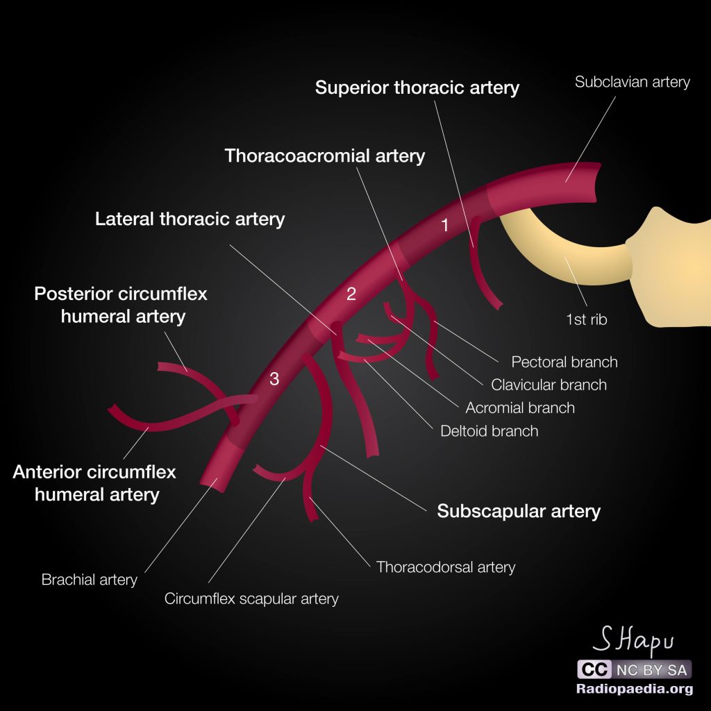
Branches of Axillary Artery

Mnemonic:
1. 1st part gives 1 branch; 2nd part gives 2 branches and 3rd part gives 3 branches.
2. Remember S AL SAP

1. 1st part: S

  • Superior thoracic artery

2. 2nd part: AL

  • Acromio-thoracic artery
  • Lateral thoracic artery

3. 3rd part: SAP

  • Subscapular artery
    • Circumflex scapular artery travels through the triangular space
  • Anterior circumflex humeral artery (ACHA)
  • Posterior circumflex humeral artery (PCHA)
    • PHCA travels through the quadrangular space

Important muscular spaces in the shoulder can be remembered using a 2 hand, 2 finger intersection analogy as described here.

Further 4 branches of acromio-thoracic artery can be remembered using the mnemonic:

Mnemonic: ABCD

  1. Acromial
  2. Breast (pectoral)
  3. Clavicular
  4. Deltoid

  • Facebook
  • Twitter
PGMEE, MRCS, USMLE, MBBS, MD/MS AnatomyCardiovascular system

Post navigation

Previous post
Next post

Related Posts

PGMEE, MRCS, USMLE, MBBS, MD/MS

Hawkin’s Classification for Talus Fractures : Mnemonic

Jun 28, 2022

Mnemonic: NISA 10-50-90-100 Type I – Non-displaced Neck fracture (upto 10% risk of AVN) 1 blood vessel damaged (entering talar neck) Management: 8 weeks in cast and 4 weeks in CAM cast Type II – Type I + Inferior (subtalar) subluxation/dislocation (upto 50% risk of AVN) 2 blood vessels damaged…

Read More
PGMEE, MRCS, USMLE, MBBS, MD/MS

Meniscal Tears on MRI

Apr 24, 2025Apr 24, 2025

Meniscal tears are best seen on T1-weighted, gradient-echo and proton-density images. The menisci are low intensity on all sequences. Morphologies Meniscal tear morphology Description MRI appearance Horizontal Separates meniscus into superior (femoral) & inferior (tibial) fragments Primarily horizontal signal on sagittal images Vertical radial Splits central margin of meniscus Vertical…

Read More
PGMEE, MRCS, USMLE, MBBS, MD/MS

Working formulation of NHL (Mnemonic)

Jan 16, 2019Feb 26, 2019

All follicular except large cell: Low grade All diffuse: Intermediate grade Blastic and non-cleaved: High grade

Read More

Leave a Reply Cancel reply

Your email address will not be published. Required fields are marked *

This site uses Akismet to reduce spam. Learn how your comment data is processed.

Pre-clinical (Basic Sciences)

Anatomy

Biochemistry

Community medicine (PSM)

Embryology

Microbiology

Pathology

Pharmacology

Physiology

Clinical Sciences

Anesthesia

Dermatology

Emergency medicine

Forensic

Internal medicine

Gynecology & Obstetrics

Oncology

Ophthalmology

Orthopedics

Otorhinolaryngology (ENT)

Pediatrics

Psychiatry

Radiology

Surgery

RSS Ask Epomedicine

  • What to study for Clinical examination in Orthopedics?
  • What is the mechanism of AVNRT?

Epomedicine weekly

  • About Epomedicine
  • Contact Us
  • Author Guidelines
  • Submit Article
  • Editorial Board
  • USMLE
  • MRCS
  • Thesis
©2026 Epomedicine | WordPress Theme by SuperbThemes